4YVN image
Deposition Date 2015-03-20
Release Date 2016-03-30
Last Version Date 2024-11-20
Entry Detail
PDB ID:
4YVN
Keywords:
Title:
Crystal structure of CotA laccase complexed with ABTS at a novel binding site
Biological Source:
Source Organism:
Host Organism:
Method Details:
Experimental Method:
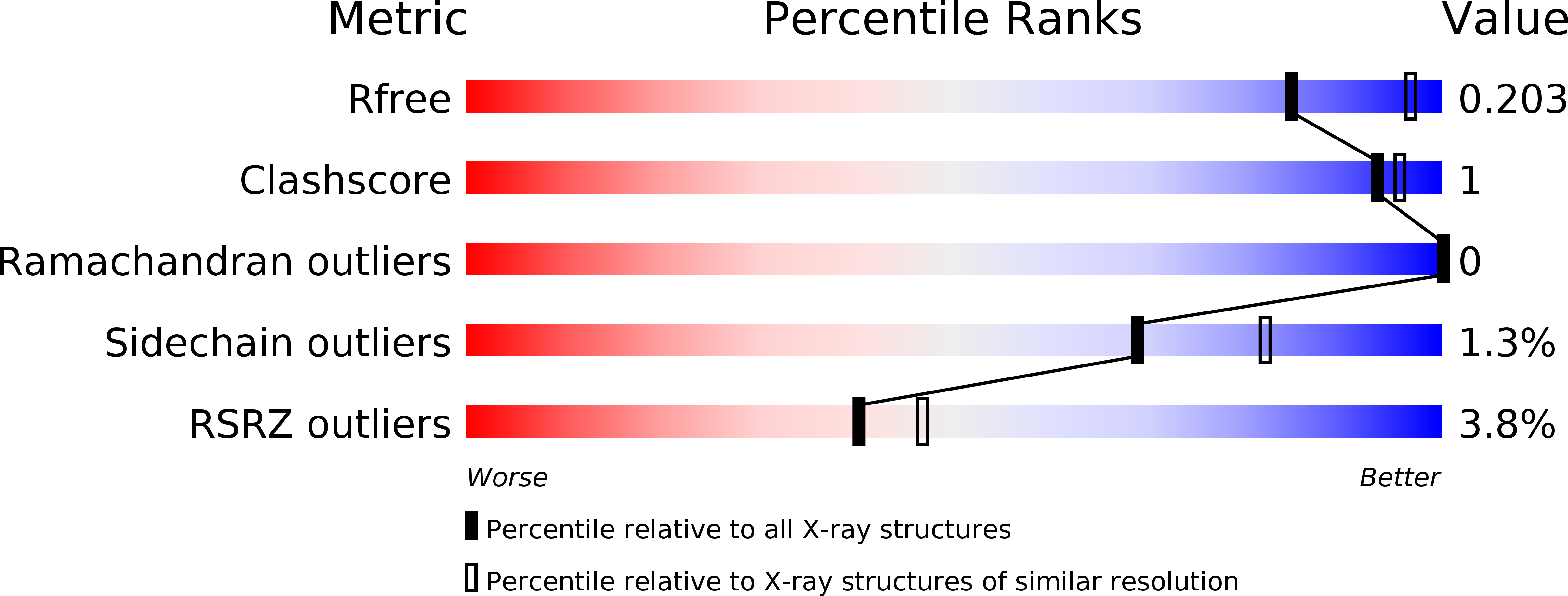
Resolution:
2.30 Å
R-Value Free:
0.19
R-Value Work:
0.15
R-Value Observed:
0.15
Space Group:
P 31 2 1
Macromolecular Entities
Structures with similar UniProt ID
Protein Blast
Polymer Type:polypeptide(L)
Molecule:Spore coat protein A
Gene (Uniprot):cotA
Chain IDs:A
Chain Length:513
Number of Molecules:1
Biological Source:Bacillus subtilis (strain 168)
Primary Citation
Crystal structure of CotA laccase complexed with 2,2-azinobis-(3-ethylbenzothiazoline-6-sulfonate) at a novel binding site
Acta Crystallogr.,Sect.F 72 328 335 (2016)
PMID: 27050268 DOI: 10.1107/S2053230X1600426X

Abstact

The CotA laccase from Bacillus subtilis is an abundant component of the spore outer coat and has been characterized as a typical laccase. The crystal structure of CotA complexed with 2,2-azinobis-(3-ethylbenzothiazoline-6-sulfonate) (ABTS) in a hole motif has been solved. The novel binding site was about 26 Å away from the T1 binding pocket. Comparison with known structures of other laccases revealed that the hole is a specific feature of CotA. The key residues Arg476 and Ser360 were directly bound to ABTS. Site-directed mutagenesis studies revealed that the residues Arg146, Arg429 and Arg476, which are located at the bottom of the novel binding site, are essential for the oxidation of ABTS and syringaldazine. Specially, a Thr480Phe variant was identified to be almost 3.5 times more specific for ABTS than for syringaldazine compared with the wild type. These results suggest this novel binding site for ABTS could be a potential target for protein engineering of CotA laccases.

Legend

Protein

Chemical

Disease

Primary Citation of related structures
Feedback Form
Name
Email
Institute
Feedback