4YV5 image
Entry Detail
PDB ID:
4YV5
Keywords:
Title:
Crystal Structure of Myotoxin II from Bothrops moojeni complexed to Suramin
Biological Source:
Source Organism:
PDB Version:
Deposition Date:
2015-03-19
Release Date:
2015-10-14
Method Details:
Experimental Method:
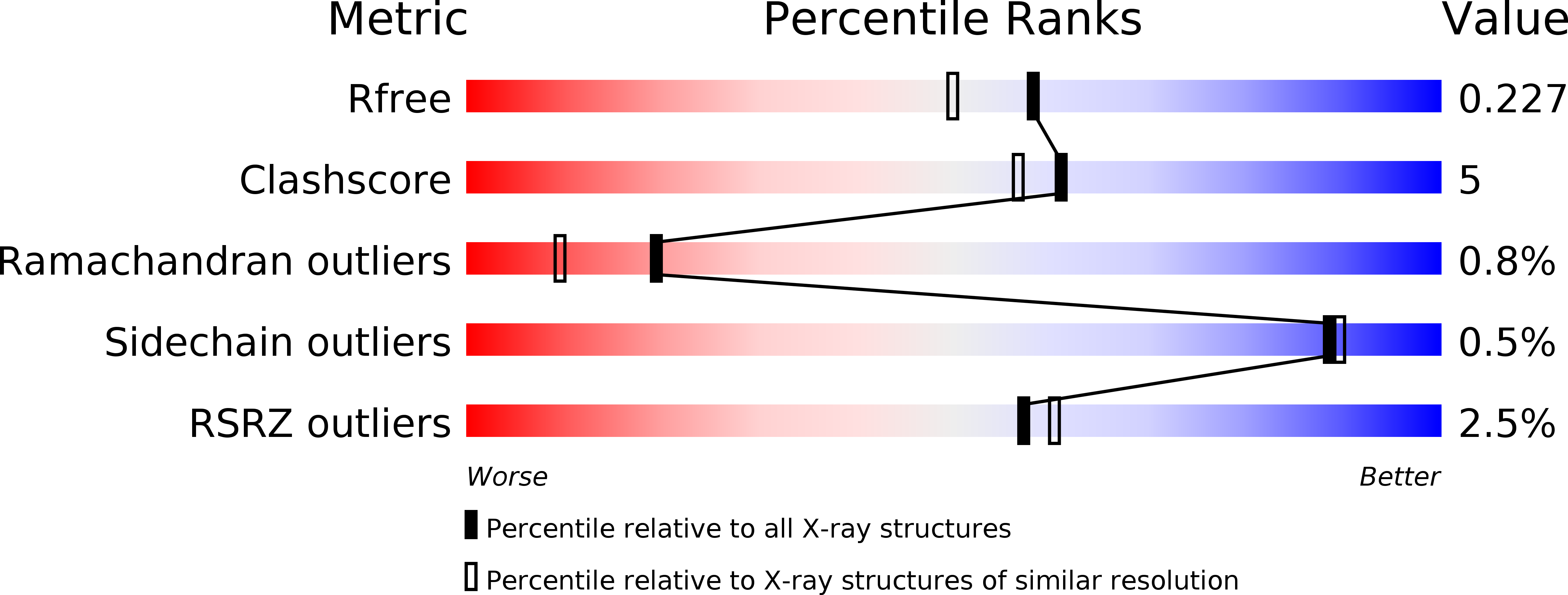
Resolution:
1.90 Å
R-Value Free:
0.22
R-Value Work:
0.19
R-Value Observed:
0.19
Space Group:
P 21 21 21
Macromolecular Entities
Polymer Type:polypeptide(L)
Description:Basic phospholipase A2 homolog 2
Chain IDs:A (auth: B), B (auth: A)
Chain Length:122
Number of Molecules:2
Biological Source:Bothrops moojeni
Primary Citation
Structural and functional evidence for membrane docking and disruption sites on phospholipase A2-like proteins revealed by complexation with the inhibitor suramin.
Acta Crystallogr. D Biol. Crystallogr. 71 2066 2078 (2015)
PMID: 26457430 DOI: 10.1107/S1399004715014443

Abstact

Local myonecrosis resulting from snakebite envenomation is not efficiently neutralized by regular antivenom administration. This limitation is considered to be a significant health problem by the World Health Organization. Phospholipase A2-like (PLA2-like) proteins are among the most important proteins related to the muscle damage resulting from several snake venoms. However, despite their conserved tertiary structure compared with PLA2s, their biological mechanism remains incompletely understood. Different oligomeric conformations and binding sites have been identified or proposed, leading to contradictory data in the literature. In the last few years, a comprehensive hypothesis has been proposed based on fatty-acid binding, allosteric changes and the presence of two different interaction sites. In the present study, a combination of techniques were used to fully understand the structural-functional characteristics of the interaction between suramin and MjTX-II (a PLA2-like toxin). In vitro neuromuscular studies were performed to characterize the biological effects of the protein-ligand interaction and demonstrated that suramin neutralizes the myotoxic activity of MjTX-II. The high-resolution structure of the complex identified the toxin-ligand interaction sites. Calorimetric assays showed two different binding events between the protein and the inhibitor. It is demonstrated for the first time that the inhibitor binds to the surface of the toxin, obstructing the sites involved in membrane docking and disruption according to the proposed myotoxic mechanism. Furthermore, higher-order oligomeric formation by interaction with interfacial suramins was observed, which may also aid the inhibitory process. These results further substantiate the current myotoxic mechanism and shed light on the search for efficient inhibitors of the local myonecrosis phenomenon.

Legend

Protein

Chemical

Disease

Primary Citation of related structures