
Deposition Date
2015-03-18
Release Date
2016-03-23
Last Version Date
2023-11-08
Entry Detail
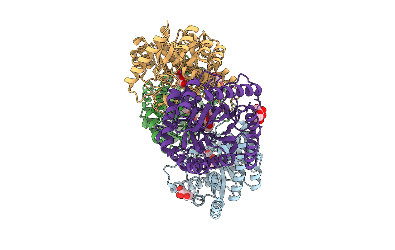
PDB ID:
4YTT
Keywords:
Title:
Crystal structure of D-tagatose 3-epimerase C66S from Pseudomonas cichorii in complex with 6-deoxy L-psicose
Biological Source:
Source Organism(s):
Pseudomonas cichorii (Taxon ID: 36746)
Expression System(s):
Method Details:
Experimental Method:
Resolution:
1.80 Å
R-Value Free:
0.22
R-Value Work:
0.19
R-Value Observed:
0.19
Space Group:
P 1 21 1
