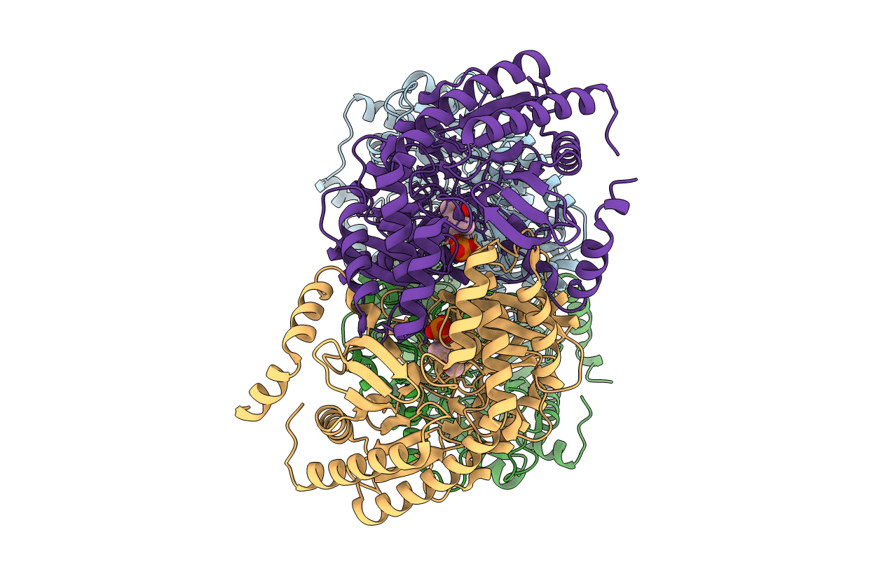
4YSN image
Entry Detail
PDB ID:
4YSN
Keywords:
Title:
Structure of aminoacid racemase in complex with PLP
Biological Source:
Source Organism:
Host Organism:
PDB Version:
Deposition Date:
2015-03-17
Release Date:
2016-04-20
Method Details:
Experimental Method:
Resolution:
1.94 Å
R-Value Free:
0.18
R-Value Work:
0.14
R-Value Observed:
0.14
Space Group:
P 21 21 21
Macromolecular Entities
Polymer Type:polypeptide(L)
Description:Putative 4-aminobutyrate aminotransferase
Chain IDs:A, B, C, D
Chain Length:462
Number of Molecules:4
Biological Source:Lactobacillus buchneri
Ligand Molecules
Primary Citation
Crystal structure of the novel amino-acid racemase isoleucine 2-epimerase from Lactobacillus buchneri.
Acta Crystallogr D Struct Biol 73 428 437 (2017)
PMID: 28471367 DOI: 10.1107/S2059798317005332

Abstact

Crystal structures of Lactobacillus buchneri isoleucine 2-epimerase, a novel branched-chain amino-acid racemase, were determined for the enzyme in the apo form, in complex with pyridoxal 5'-phosphate (PLP), in complex with N-(5'-phosphopyridoxyl)-L-isoleucine (PLP-L-Ile) and in complex with N-(5'-phosphopyridoxyl)-D-allo-isoleucine (PLP-D-allo-Ile) at resolutions of 2.77, 1.94, 2.65 and 2.12 Å, respectively. The enzyme assembled as a tetramer, with each subunit being composed of N-terminal, C-terminal and large PLP-binding domains. The active-site cavity in the apo structure was much more solvent-accessible than that in the PLP-bound structure. This indicates that a marked structural change occurs around the active site upon binding of PLP that provides a solvent-inaccessible environment for the enzymatic reaction. The main-chain coordinates of the L. buchneri isoleucine 2-epimerase monomer showed a notable similarity to those of α-amino-ℇ-caprolactam racemase from Achromobactor obae and γ-aminobutyrate aminotransferase from Escherichia coli. However, the amino-acid residues involved in substrate binding in those two enzymes are only partially conserved in L. buchneri isoleucine 2-epimerase, which may account for the differences in substrate recognition by the three enzymes. The structures bound with reaction-intermediate analogues (PLP-L-Ile and PLP-D-allo-Ile) and site-directed mutagenesis suggest that L-isoleucine epimerization proceeds through abstraction of the α-hydrogen of the substrate by Lys280, while Asp222 serves as the catalytic residue adding an α-hydrogen to the quinonoid intermediate to form D-allo-isoleucine.

Legend

Protein

Chemical

Disease

Primary Citation of related structures