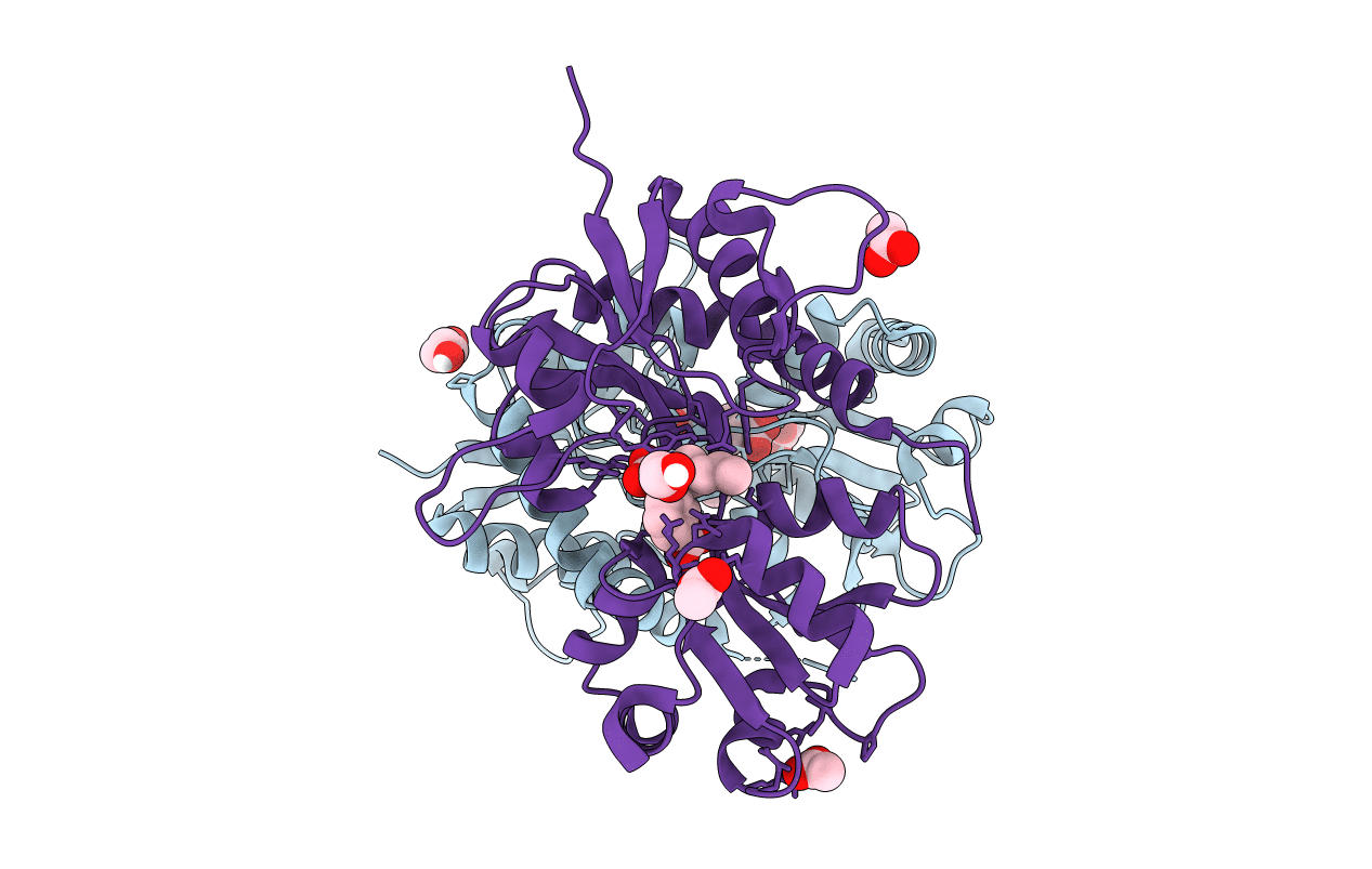
4YMB image
Deposition Date 2015-03-06
Release Date 2015-08-05
Last Version Date 2024-11-06
Entry Detail
PDB ID:
4YMB
Title:
Structure of the ligand-binding domain of GluK1 in complex with the antagonist CNG10111
Biological Source:
Source Organism:
Host Organism:
Method Details:
Experimental Method:
Resolution:
1.93 Å
R-Value Free:
0.21
R-Value Work:
0.17
R-Value Observed:
0.18
Space Group:
P 41 21 2
Macromolecular Entities
Polymer Type:polypeptide(L)
Molecule:Glutamate receptor ionotropic, kainate 1,Glutamate receptor ionotropic, kainate 1
Gene (Uniprot):Grik1
Chain IDs:A, B
Chain Length:257
Number of Molecules:2
Biological Source:Rattus norvegicus
Primary Citation
Structure-Activity Relationship Study of Ionotropic Glutamate Receptor Antagonist (2S,3R)-3-(3-Carboxyphenyl)pyrrolidine-2-carboxylic Acid.
J.Med.Chem. 58 6131 6150 (2015)
PMID: 26200741 DOI: 10.1021/acs.jmedchem.5b00750

Abstact

Herein we describe the first structure-activity relationship study of the broad-range iGluR antagonist (2S,3R)-3-(3-carboxyphenyl)pyrrolidine-2-carboxylic acid (1) by exploring the pharmacological effect of substituents in the 4, 4', or 5' positions and the bioisosteric substitution of the distal carboxylic acid for a phosphonic acid moiety. Of particular interest is a hydroxyl group in the 4' position 2a which induced a preference in binding affinity for homomeric GluK3 over GluK1 (Ki = 0.87 and 4.8 μM, respectively). Two X-ray structures of ligand binding domains were obtained: 2e in GluA2-LBD and 2f in GluK1-LBD, both at 1.9 Å resolution. Compound 2e induces a D1-D2 domain opening in GluA2-LBD of 17.3-18.8° and 2f a domain opening in GluK1-LBD of 17.0-17.5° relative to the structures with glutamate. The pyrrolidine-2-carboxylate moiety of 2e and 2f shows a similar binding mode as kainate. The 3-carboxyphenyl ring of 2e and 2f forms contacts comparable to those of the distal carboxylate in kainate.

Legend

Protein

Chemical

Disease

Primary Citation of related structures