4YLB image
Deposition Date 2015-03-05
Release Date 2015-11-04
Last Version Date 2024-03-20
Entry Detail
PDB ID:
4YLB
Keywords:
Title:
Crystal Structure of A102D mutant of hsp14.1 from Sulfolobus solfatataricus P2
Biological Source:
Source Organism(s):
Expression System(s):
Method Details:
Experimental Method:
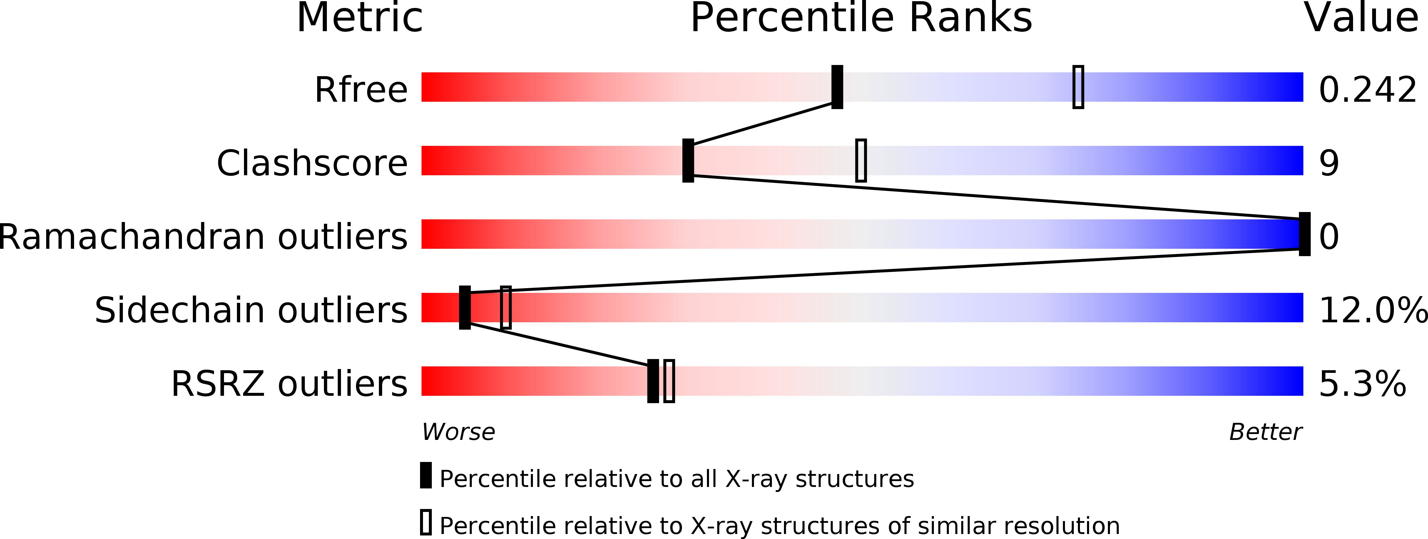
Resolution:
2.50 Å
R-Value Free:
0.24
R-Value Work:
0.19
R-Value Observed:
0.19
Space Group:
H 3 2
Macromolecular Entities
Structures with similar UniProt ID
Protein Blast
Polymer Type:polypeptide(L)
Molecule:Heat shock protein Hsp20
Gene (Uniprot):Ssol_0413
Mutagens:A102D
Chain IDs:A, B, C, D
Chain Length:128
Number of Molecules:4
Biological Source:Sulfolobus solfataricus (strain 98/2)
Ligand Molecules
Primary Citation
Active-State Structures of a Small Heat-Shock Protein Revealed a Molecular Switch for Chaperone Function
Structure 23 2066 2075 (2015)
PMID: 26439766 DOI: 10.1016/j.str.2015.08.015

Abstact

Small heat-shock proteins (sHsps) maintain cellular homeostasis by binding to denatured client proteins to prevent aggregation. Numerous studies indicate that the N-terminal domain (NTD) of sHsps is responsible for binding to client proteins, but the binding mechanism and chaperone activity regulation remain elusive. Here, we report the crystal structures of the wild-type and mutants of an sHsp from Sulfolobus solfataricus representing the inactive and active state of this protein, respectively. All three structures reveal well-defined NTD, but their conformations are remarkably different. The mutant NTDs show disrupted helices presenting a reformed hydrophobic surface compatible with recognizing client proteins. Our functional data show that mutating key hydrophobic residues in this region drastically altered the chaperone activity of this sHsp. These data suggest a new model in which a molecular switch located in NTD facilitates conformational changes for client protein binding.

Legend

Protein

Chemical

Disease

Primary Citation of related structures
Feedback Form
Name
Email
Institute
Feedback