4YJ6 image
Deposition Date 2015-03-03
Release Date 2015-11-04
Last Version Date 2024-11-13
Entry Detail
PDB ID:
4YJ6
Keywords:
Title:
The Crystal Structure of a Bacterial Aryl Acylamidase Belonging to the Amidase signature (AS) enzymes family
Biological Source:
Source Organism:
Method Details:
Experimental Method:
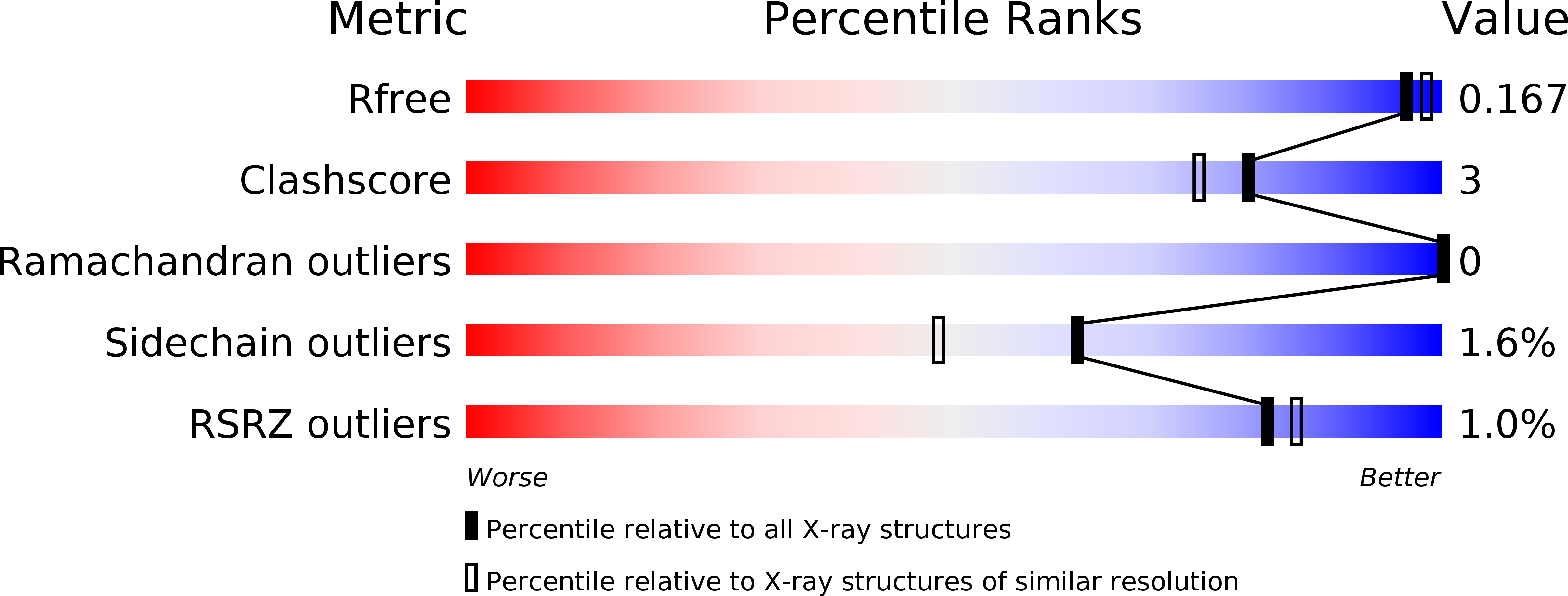
Resolution:
1.70 Å
R-Value Free:
0.16
R-Value Work:
0.15
R-Value Observed:
0.15
Space Group:
P 41 21 2
Macromolecular Entities
Structures with similar UniProt ID
Protein Blast
Polymer Type:polypeptide(L)
Molecule:Aryl acylamidase
Chain IDs:A
Chain Length:501
Number of Molecules:1
Biological Source:bacterium CSBL00001
Modified Residue
Compound ID Chain ID Parent Comp ID Details 2D Image
MSE A MET modified residue
Ligand Molecules
Primary Citation
Crystal structure analysis of a bacterial aryl acylamidase belonging to the amidase signature enzyme family
Biochem.Biophys.Res.Commun. 467 268 274 (2015)
PMID: 26454172 DOI: 10.1016/j.bbrc.2015.09.177

Abstact

The atomic structure of a bacterial aryl acylamidase (EC 3.5.1.13; AAA) is reported and structural features are investigated to better understand the catalytic profile of this enzyme. Structures of AAA were determined in its native form and in complex with the analgesic acetanilide, p-acetaminophenol, at 1.70 Å and 1.73 Å resolutions, respectively. The overall structural fold of AAA was identified as an α/β fold class, exhibiting an open twisted β-sheet core surrounded by α-helices. The asymmetric unit contains one AAA molecule and the monomeric form is functionally active. The core structure enclosing the signature sequence region, including the canonical Ser-cisSer-Lys catalytic triad, is conserved in all members of the Amidase Signature enzyme family. The structure of AAA in a complex with its ligand reveals a unique organization in the substrate-binding pocket. The binding pocket consists of two loops (loop1 and loop2) in the amidase signature sequence and one helix (α10) in the non-amidase signature sequence. We identified two residues (Tyr(136) and Thr(330)) that interact with the ligand via water molecules, and a hydrogen-bonding network that explains the catalytic affinity over various aryl acyl compounds. The optimum activity of AAA at pH > 10 suggests that the reaction mechanism employs Lys(84) as the catalytic base to polarize the Ser(187) nucleophile in the catalytic triad.

Legend

Protein

Chemical

Disease

Primary Citation of related structures
Feedback Form
Name
Email
Institute
Feedback