Deposition Date
2015-02-28
Release Date
2015-12-02
Last Version Date
2023-09-27
Entry Detail
PDB ID:
4YI9
Keywords:
Title:
Crystal structure of non-myristoylated E153A recoverin at 1.35 A resolution with a sodium ion bound to EF-hand 2 and calcium ion bound to EF-hand 3
Biological Source:
Source Organism(s):
Bos taurus (Taxon ID: 9913)
Expression System(s):
Method Details:
Experimental Method:
Resolution:
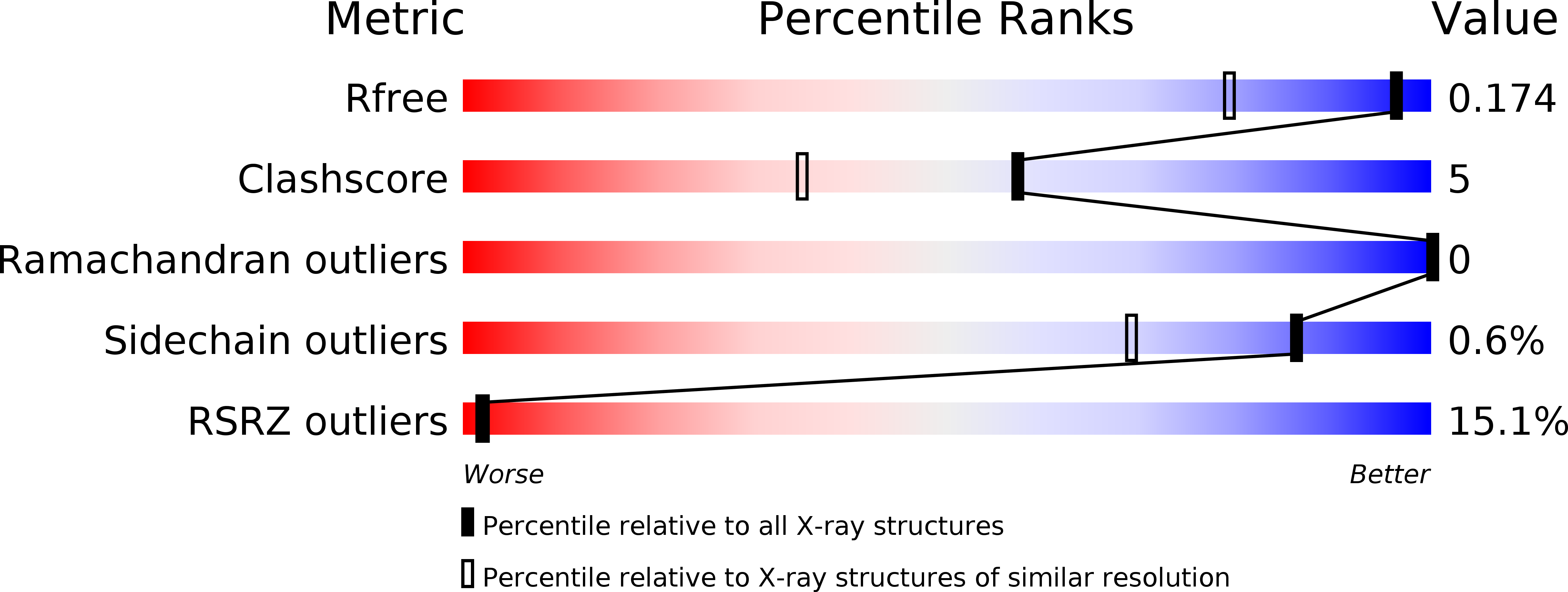
1.35 Å
R-Value Free:
0.17
R-Value Work:
0.15
R-Value Observed:
0.15
Space Group:
I 4