
Deposition Date
2015-02-26
Release Date
2015-04-22
Last Version Date
2024-03-20
Entry Detail
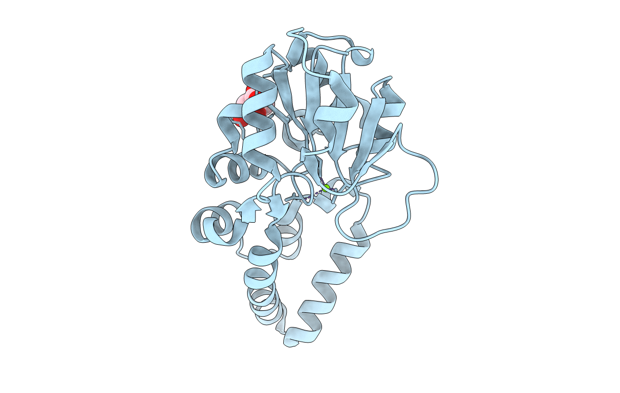
PDB ID:
4YGS
Keywords:
Title:
Crystal structure of HAD phosphatase from Thermococcus onnurineus
Biological Source:
Source Organism(s):
Thermococcus onnurineus (strain NA1) (Taxon ID: 523850)
Expression System(s):
Method Details:
Experimental Method:
Resolution:
1.70 Å
R-Value Free:
0.19
R-Value Work:
0.15
R-Value Observed:
0.15
Space Group:
C 1 2 1
