4YG2 image
Deposition Date 2015-02-25
Release Date 2015-03-18
Last Version Date 2024-02-28
Entry Detail
PDB ID:
4YG2
Title:
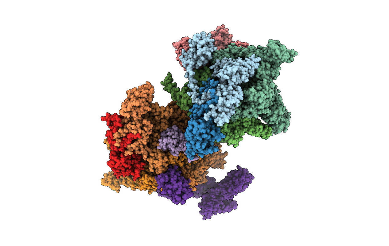
X-ray crystal structur of Escherichia coli RNA polymerase sigma70 holoenzyme
Biological Source:
Source Organism:
Host Organism:
Method Details:
Experimental Method:
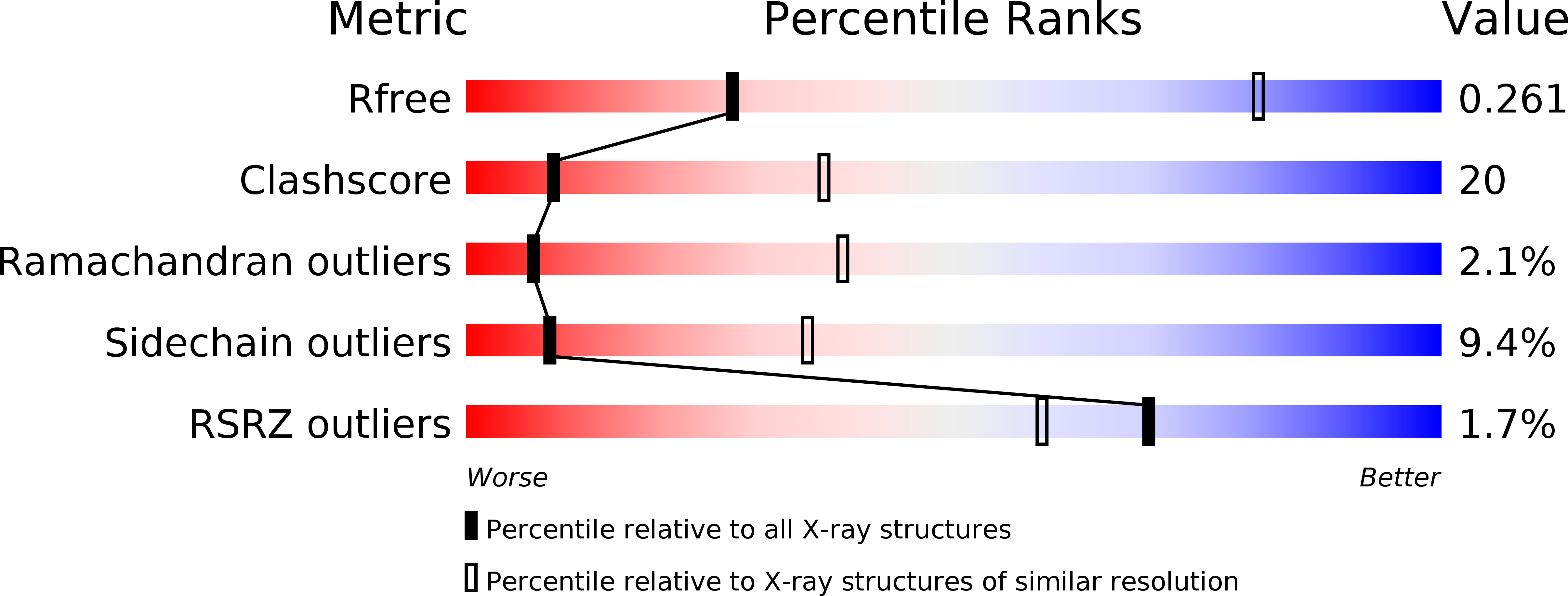
Resolution:
3.70 Å
R-Value Free:
0.26
R-Value Work:
0.19
R-Value Observed:
0.19
Space Group:
P 21 21 21
Macromolecular Entities
Structures with similar UniProt ID
Protein Blast
Polymer Type:polypeptide(L)
Molecule:DNA-directed RNA polymerase subunit alpha
Gene (Uniprot):rpoA
Chain IDs:A, B, G, H
Chain Length:329
Number of Molecules:4
Biological Source:Escherichia coli O157:H7
Structures with similar UniProt ID
Protein Blast
Polymer Type:polypeptide(L)
Molecule:DNA-directed RNA polymerase subunit beta
Gene (Uniprot):rpoB
Chain IDs:C, I
Chain Length:1342
Number of Molecules:2
Biological Source:Escherichia coli O157:H7
Structures with similar UniProt ID
Protein Blast
Polymer Type:polypeptide(L)
Molecule:DNA-directed RNA polymerase subunit beta'
Gene (Uniprot):rpoC
Chain IDs:D, J
Chain Length:1407
Number of Molecules:2
Biological Source:Escherichia coli O157:H7
Structures with similar UniProt ID
Protein Blast
Polymer Type:polypeptide(L)
Molecule:DNA-directed RNA polymerase subunit omega
Gene (Uniprot):rpoZ
Chain IDs:E, K
Chain Length:91
Number of Molecules:2
Biological Source:Escherichia coli O157:H7
Structures with similar UniProt ID
Protein Blast
Polymer Type:polypeptide(L)
Molecule:RNA polymerase sigma factor RpoD
Gene (Uniprot):rpoD
Chain IDs:F, L
Chain Length:613
Number of Molecules:2
Biological Source:Escherichia coli K12
Primary Citation
X-ray crystal structure of Escherichia coli RNA polymerase sigma70 holoenzyme
J. Biol. Chem. 288 9126 9134 (2013)
PMID: 23389035 DOI: 10.1074/jbc.M112.430900

Abstact

Escherichia coli RNA polymerase (RNAP) is the most studied bacterial RNAP and has been used as the model RNAP for screening and evaluating potential RNAP-targeting antibiotics. However, the x-ray crystal structure of E. coli RNAP has been limited to individual domains. Here, I report the x-ray structure of the E. coli RNAP σ(70) holoenzyme, which shows σ region 1.1 (σ1.1) and the α subunit C-terminal domain for the first time in the context of an intact RNAP. σ1.1 is positioned at the RNAP DNA-binding channel and completely blocks DNA entry to the RNAP active site. The structure reveals that σ1.1 contains a basic patch on its surface, which may play an important role in DNA interaction to facilitate open promoter complex formation. The α subunit C-terminal domain is positioned next to σ domain 4 with a fully stretched linker between the N- and C-terminal domains. E. coli RNAP crystals can be prepared from a convenient overexpression system, allowing further structural studies of bacterial RNAP mutants, including functionally deficient and antibiotic-resistant RNAPs.

Legend

Protein

Chemical

Disease

Primary Citation of related structures