4YFV image
Entry Detail
PDB ID:
4YFV
Keywords:
Title:
X-ray structure of the 4-N-formyltransferase VioF from Providencia alcalifaciens O30
Biological Source:
Source Organism:
Host Organism:
PDB Version:
Deposition Date:
2015-02-25
Release Date:
2015-03-11
Method Details:
Experimental Method:
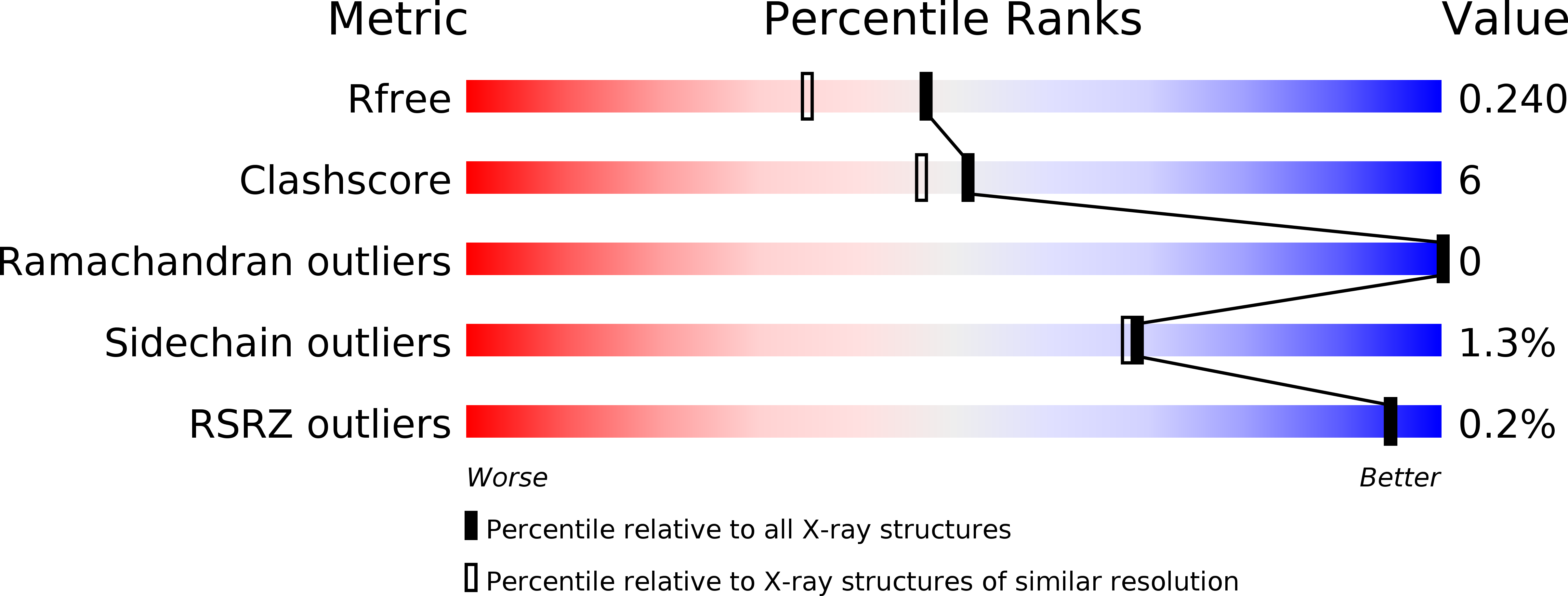
Resolution:
1.89 Å
R-Value Free:
0.23
R-Value Work:
0.18
R-Value Observed:
0.18
Space Group:
H 3 2
Macromolecular Entities
Polymer Type:polypeptide(L)
Description:VioF
Chain IDs:A, B
Chain Length:254
Number of Molecules:2
Biological Source:Providencia alcalifaciens
Primary Citation
Molecular structure of an N-formyltransferase from Providencia alcalifaciens O30.
Protein Sci. 24 976 986 (2015)
PMID: 25752909 DOI: 10.1002/pro.2675

Abstact

The existence of N-formylated sugars in the O-antigens of Gram-negative bacteria has been known since the middle 1980s, but only recently have the biosynthetic pathways for their production been reported. In these pathways, glucose-1-phosphate is first activated by attachment to a dTMP moiety. This step is followed by a dehydration reaction and an amination. The last step in these pathways is catalyzed by N-formyltransferases that utilize N(10) -formyltetrahydrofolate as the carbon source. Here we describe the three-dimensional structure of one of these N-formyltransferases, namely VioF from Providencia alcalifaciens O30. Specifically, this enzyme catalyzes the conversion of dTDP-4-amino-4,6-dideoxyglucose (dTDP-Qui4N) to dTDP-4,6-dideoxy-4-formamido-d-glucose (dTDP-Qui4NFo). For this analysis, the structure of VioF was solved to 1.9 Å resolution in both its apoform and in complex with tetrahydrofolate and dTDP-Qui4N. The crystals used in the investigation belonged to the space group R32 and demonstrated reticular merohedral twinning. The overall catalytic core of the VioF subunit is characterized by a six stranded mixed β-sheet flanked on one side by three α-helices and on the other side by mostly random coil. This N-terminal domain is followed by an α-helix and a β-hairpin that form the subunit:subunit interface. The active site of the enzyme is shallow and solvent-exposed. Notably, the pyranosyl moiety of dTDP-Qui4N is positioned into the active site by only one hydrogen bond provided by Lys 77. Comparison of the VioF model to that of a previously determined N-formyltransferase suggests that substrate specificity is determined by interactions between the protein and the pyrophosphoryl group of the dTDP-sugar substrate.

Legend

Protein

Chemical

Disease

Primary Citation of related structures