Deposition Date
2015-02-24
Release Date
2015-03-11
Last Version Date
2024-11-13
Entry Detail
PDB ID:
4YF2
Keywords:
Title:
Crystal structure of mouse sperm C-type lysozyme-like protein 1
Biological Source:
Source Organism(s):
Mus musculus (Taxon ID: 10090)
Expression System(s):
Method Details:
Experimental Method:
Resolution:
2.15 Å
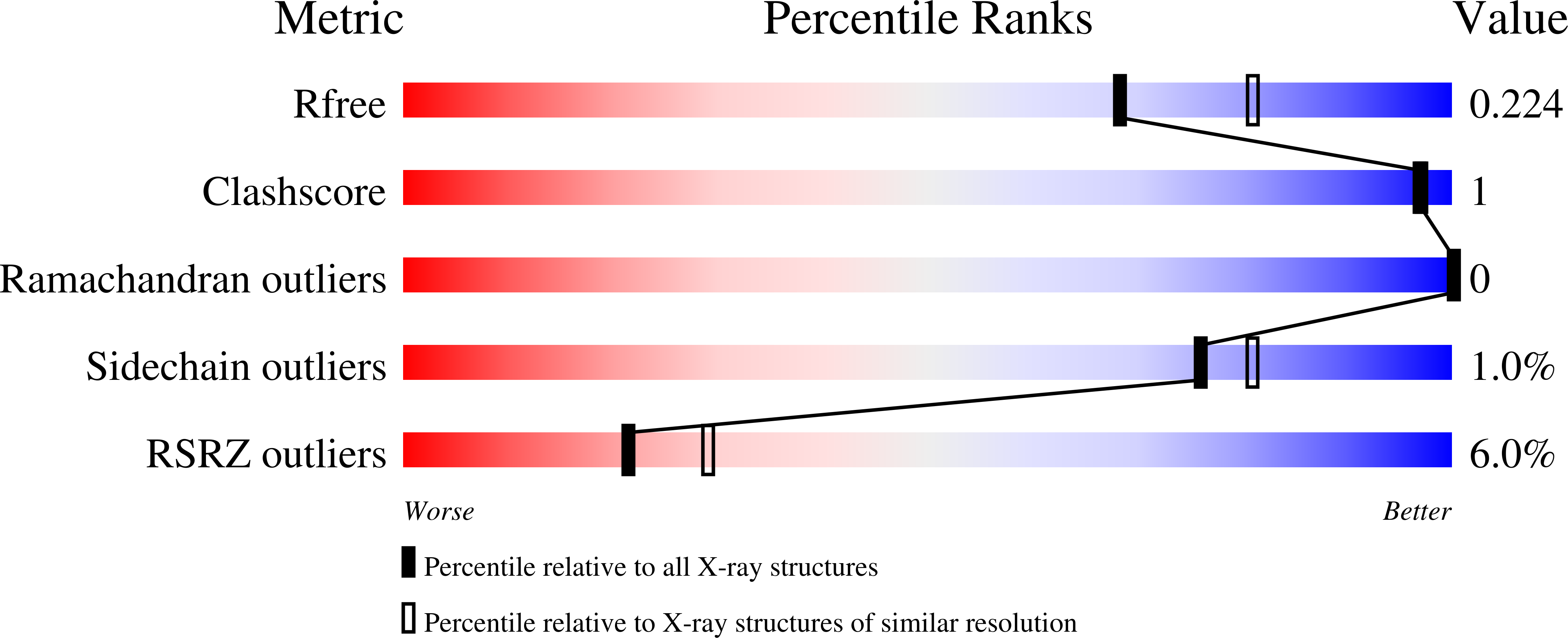
R-Value Free:
0.21
R-Value Work:
0.18
R-Value Observed:
0.18
Space Group:
P 65