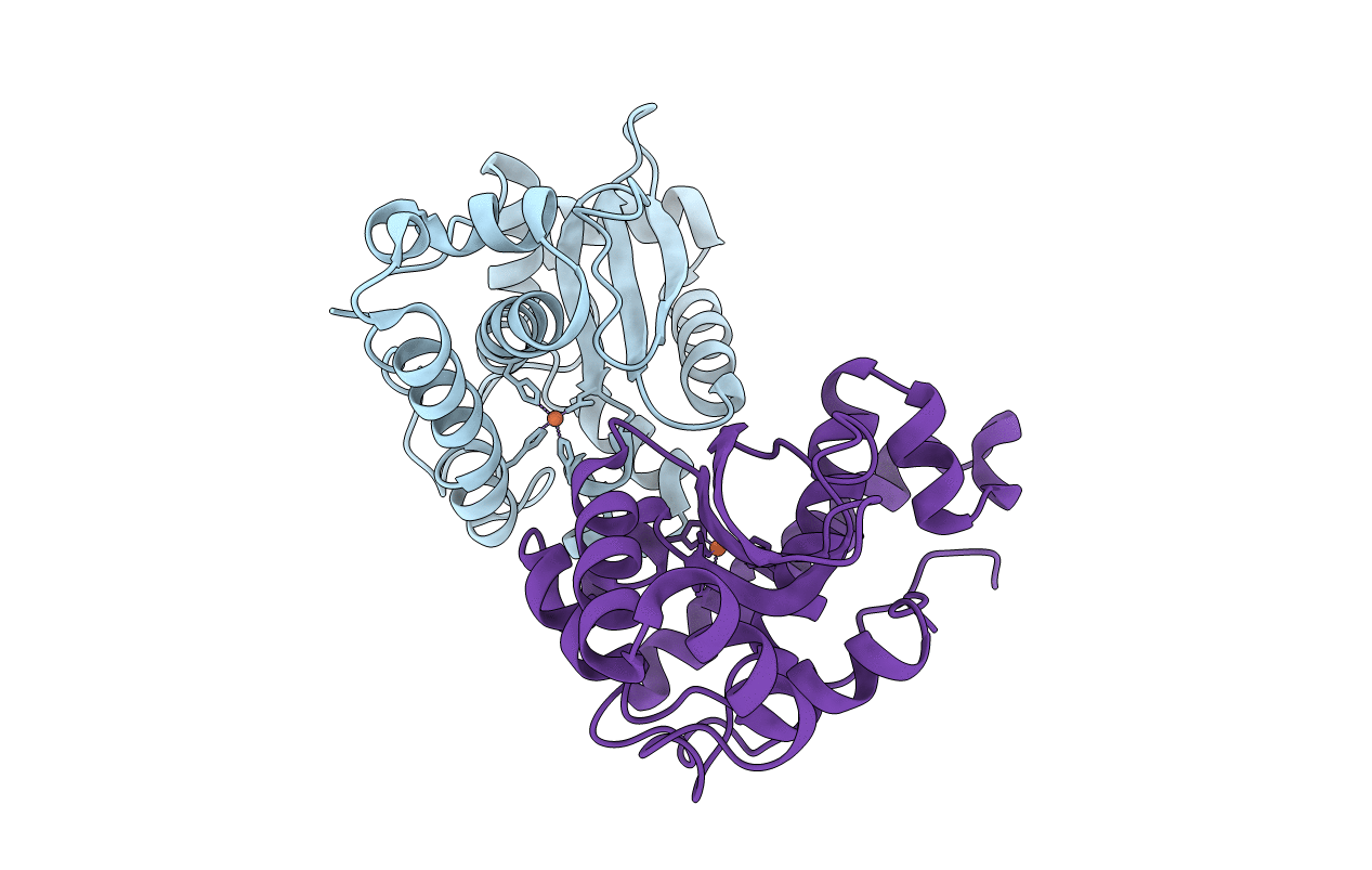
4YET image
Entry Detail
PDB ID:
4YET
Keywords:
Title:
X-ray crystal structure of superoxide dismutase from Babesia bovis solved by Sulfur SAD
Biological Source:
Source Organism:
Host Organism:
PDB Version:
Deposition Date:
2015-02-24
Release Date:
2015-04-15
Method Details:
Experimental Method:
Resolution:
1.75 Å
R-Value Free:
0.17
R-Value Work:
0.14
R-Value Observed:
0.14
Space Group:
P 21 21 21
Macromolecular Entities
Polymer Type:polypeptide(L)
Description:Superoxide dismutase
Chain IDs:A, B
Chain Length:203
Number of Molecules:2
Biological Source:Babesia bovis
Ligand Molecules
Primary Citation
Iron superoxide dismutases in eukaryotic pathogens: new insights from Apicomplexa and Trypanosoma structures.
Acta Crystallogr.,Sect.F 71 615 621 (2015)
PMID: 25961325 DOI: 10.1107/S2053230X15004185

Abstact

Prior studies have highlighted the potential of superoxide dismutases as drug targets in eukaryotic pathogens. This report presents the structures of three iron-dependent superoxide dismutases (FeSODs) from Trypanosoma cruzi, Leishmania major and Babesia bovis. Comparison with existing structures from Plasmodium and other trypanosome isoforms shows a very conserved overall fold with subtle differences. In particular, structural data suggest that B. bovis FeSOD may display similar resistance to peroxynitrite-mediated inactivation via an intramolecular electron-transfer pathway as previously described in T. cruzi FeSOD isoform B, thus providing valuable information for structure-based drug design. Furthermore, lysine-acetylation results in T. cruzi indicate that acetylation occurs at a position close to that responsible for the regulation of acetylation-mediated activity in the human enzyme.

Legend

Protein

Chemical

Disease

Primary Citation of related structures