4YD2 image
Deposition Date 2015-02-20
Release Date 2015-08-05
Last Version Date 2023-09-27
Entry Detail
PDB ID:
4YD2
Keywords:
Title:
Nicked complex of human DNA Polymerase Mu with 2-nt gapped DNA substrate
Biological Source:
Source Organism:
Homo sapiens (Taxon ID: 9606)
Host Organism:
Method Details:
Experimental Method:
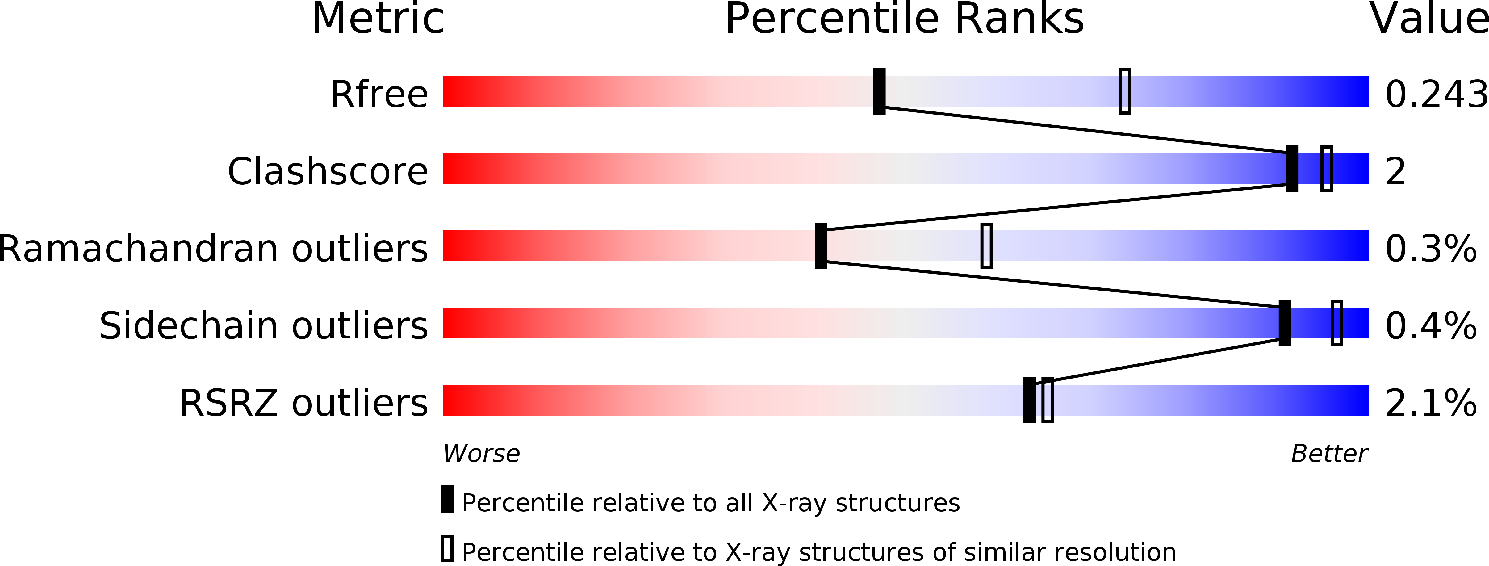
Resolution:
2.47 Å
R-Value Free:
0.24
R-Value Work:
0.21
R-Value Observed:
0.21
Space Group:
P 21 21 21
Macromolecular Entities
Polymer Type:polypeptide(L)
Molecule:DNA-directed DNA/RNA polymerase mu
Gene (Uniprot):POLM
Mutations:deletion of residues P398-P410, replaced by glycine (labelled G410)
Chain IDs:A
Chain Length:354
Number of Molecules:1
Biological Source:Homo sapiens
Polymer Type:polydeoxyribonucleotide
Molecule:DNA (5'-D(P*GP*CP*CP*G)-3')
Chain IDs:D
Chain Length:4
Number of Molecules:1
Biological Source:Homo sapiens
Polymer Type:polydeoxyribonucleotide
Molecule:DNA (5'-D(*CP*GP*TP*AP*T)-3')
Chain IDs:C (auth: P)
Chain Length:5
Number of Molecules:1
Biological Source:Homo sapiens
Polymer Type:polydeoxyribonucleotide
Molecule:DNA (5'-D(*CP*GP*GP*CP*AP*AP*T)-3')
Chain IDs:B (auth: T)
Chain Length:10
Number of Molecules:1
Biological Source:Homo sapiens
Primary Citation
Creative template-dependent synthesis by human polymerase mu.
Proc.Natl.Acad.Sci.USA 112 E4530 E4536 (2015)
PMID: 26240373 DOI: 10.1073/pnas.1505798112

Abstact

Among the many proteins used to repair DNA double-strand breaks by nonhomologous end joining (NHEJ) are two related family X DNA polymerases, Pol λ and Pol µ. Which of these two polymerases is preferentially used for filling DNA gaps during NHEJ partly depends on sequence complementarity at the break, with Pol λ and Pol µ repairing complementary and noncomplementary ends, respectively. To better understand these substrate preferences, we present crystal structures of Pol µ on a 2-nt gapped DNA substrate, representing three steps of the catalytic cycle. In striking contrast to Pol λ, Pol µ "skips" the first available template nucleotide, instead using the template base at the 5' end of the gap to direct nucleotide binding and incorporation. This remarkable divergence from canonical 3'-end gap filling is consistent with data on end-joining substrate specificity in cells, and provides insights into polymerase substrate choices during NHEJ.

Legend

Protein

Chemical

Disease

Primary Citation of related structures