4Y89 image
Deposition Date 2015-02-16
Release Date 2015-09-02
Last Version Date 2023-09-27
Entry Detail
PDB ID:
4Y89
Keywords:
Title:
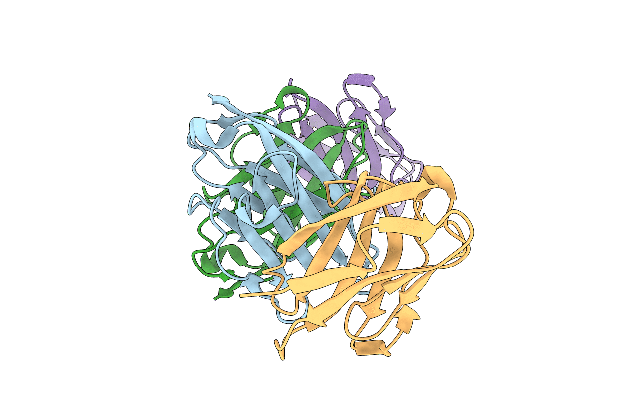
Crystal structure of the N-terminal domain of CEACAM7
Biological Source:
Source Organism:
Homo sapiens (Taxon ID: 9606)
Method Details:
Experimental Method:
Resolution:
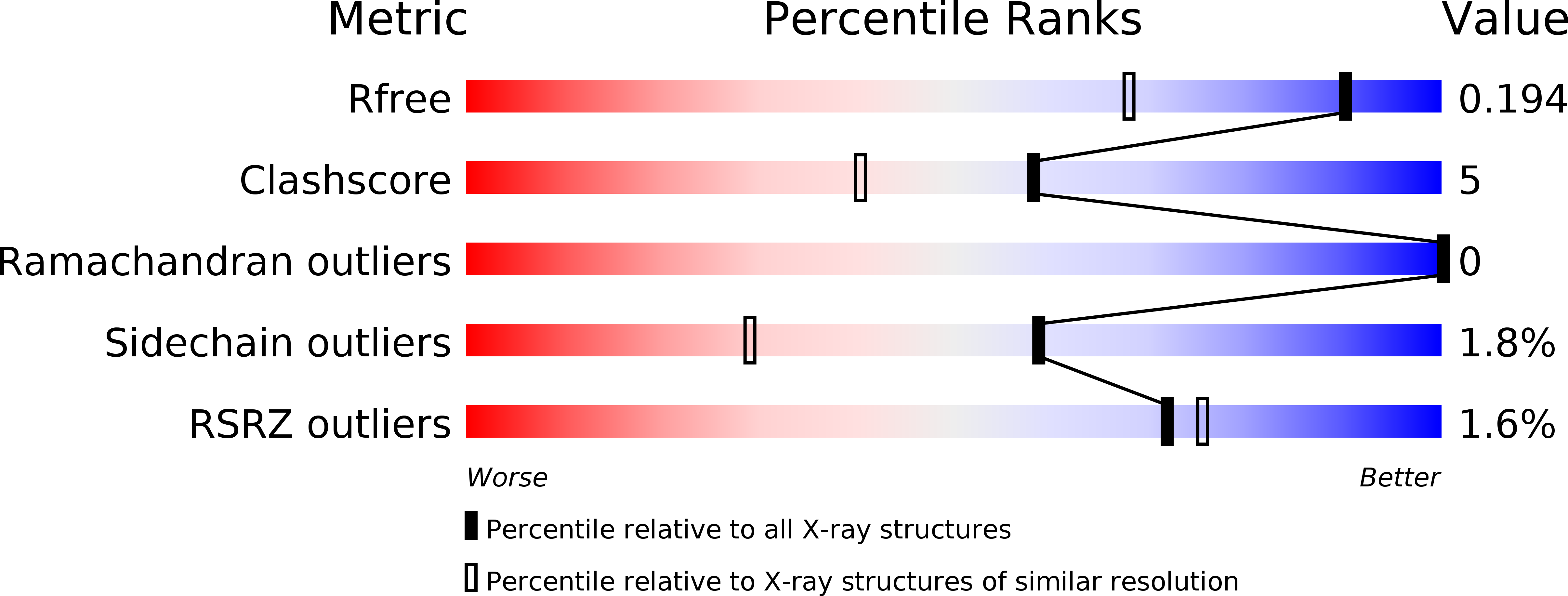
1.47 Å
R-Value Free:
0.19
R-Value Work:
0.14
R-Value Observed:
0.14
Space Group:
P 1 21 1
Macromolecular Entities
Structures with similar UniProt ID
Protein Blast
Polymer Type:polypeptide(L)
Molecule:Carcinoembryonic antigen-related cell adhesion molecule 7
Chain IDs:A, B, C, D
Chain Length:109
Number of Molecules:4
Biological Source:Homo sapiens
Ligand Molecules
Primary Citation
Structure of the N-terminal dimerization domain of CEACAM7.
Acta Crystallogr.,Sect.F 71 1169 1175 (2015)
PMID: 26323304 DOI: 10.1107/S2053230X15013576

Abstact

CEACAM7 is a human cellular adhesion protein that is expressed on the surface of colon and rectum epithelial cells and is downregulated in colorectal cancers. It achieves cell adhesion through dimerization of the N-terminal IgV domain. The crystal structure of the N-terminal dimerization domain of CEACAM has been determined at 1.47 Å resolution. The overall fold of CEACAM7 is similar to those of CEACAM1 and CEACAM5; however, there are differences, the most notable of which is an insertion that causes the C'' strand to buckle, leading to the creation of a hydrogen bond in the dimerization interface. The Kdimerization for CEACAM7 determined by sedimentation equilibrium is tenfold tighter than that measured for CEACAM5. These findings suggest that the dimerization affinities of CEACAMs are modulated via sequence variation in the dimerization surface.

Legend

Protein

Chemical

Disease

Primary Citation of related structures