4Y7M image
Deposition Date 2015-02-15
Release Date 2015-08-05
Last Version Date 2024-11-13
Entry Detail
PDB ID:
4Y7M
Title:
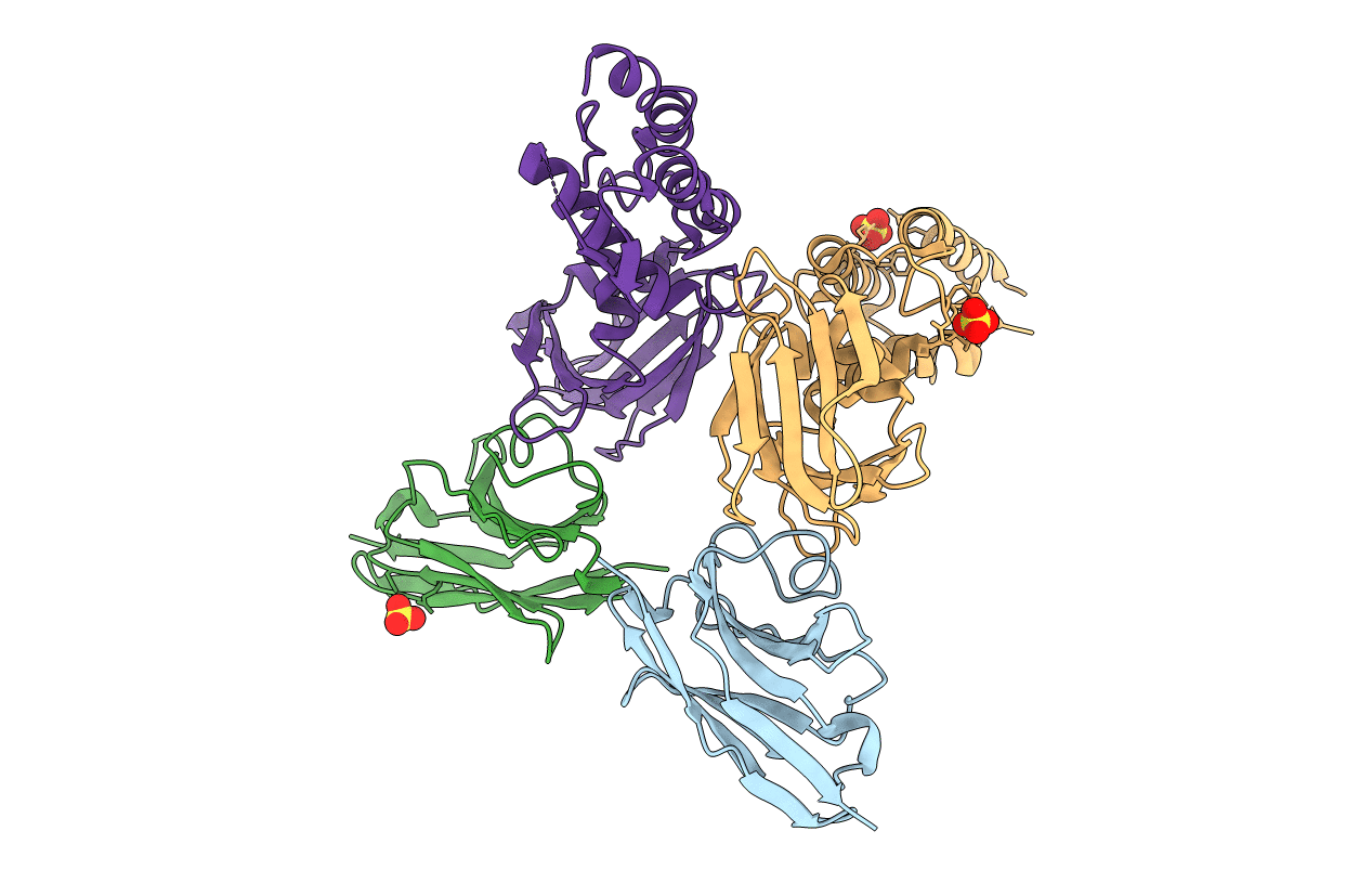
T6SS protein TssM C-terminal domain (835-1129) from EAEC
Biological Source:
Source Organism:
Method Details:
Experimental Method:
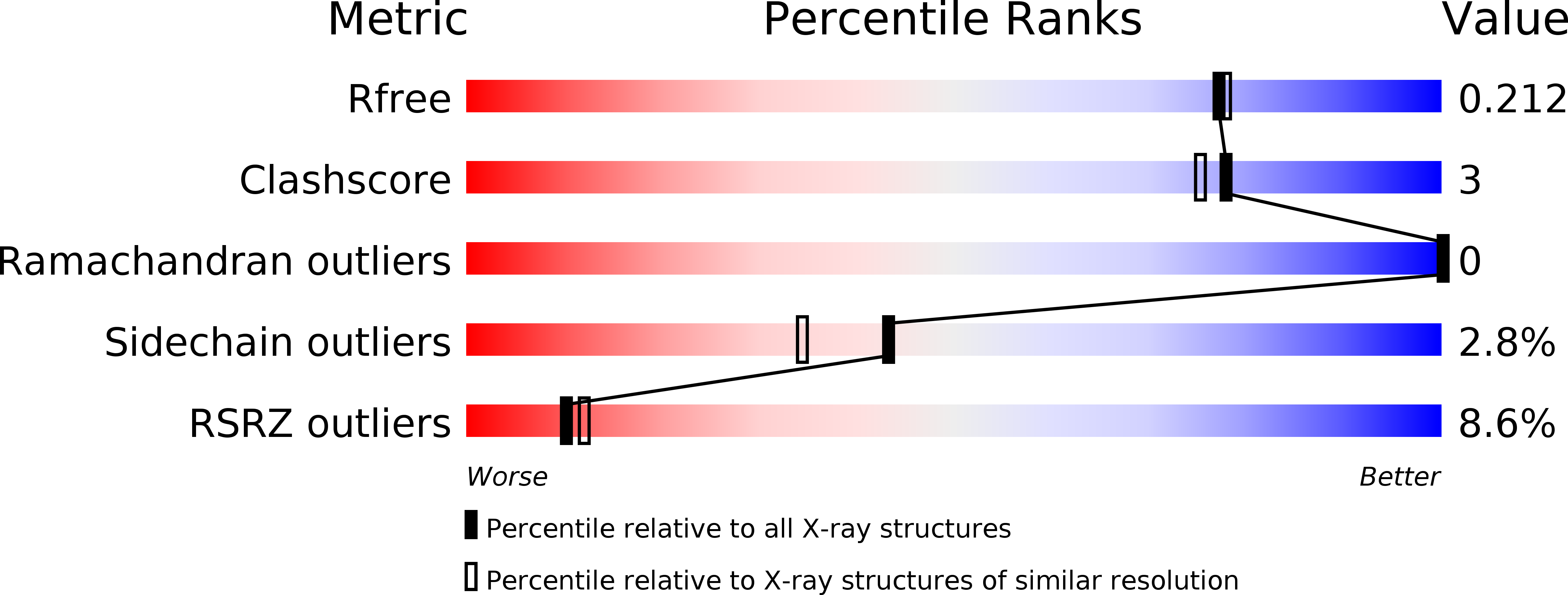
Resolution:
1.92 Å
R-Value Free:
0.21
R-Value Work:
0.18
R-Value Observed:
0.18
Space Group:
P 64
Macromolecular Entities
Polymer Type:polypeptide(L)
Molecule:Hi113 protein
Chain IDs:A, B
Chain Length:129
Number of Molecules:2
Biological Source:Lama glama
Polymer Type:polypeptide(L)
Molecule:Type VI secretion protein IcmF
Chain IDs:C, D
Chain Length:295
Number of Molecules:2
Biological Source:Escherichia coli 2-156-04_S3_C3
Ligand Molecules
Primary Citation
Biogenesis and structure of a type VI secretion membrane core complex.
Nature 523 555 560 (2015)
PMID: 26200339 DOI: 10.1038/nature14667

Abstact

Bacteria share their ecological niches with other microbes. The bacterial type VI secretion system is one of the key players in microbial competition, as well as being an important virulence determinant during bacterial infections. It assembles a nano-crossbow-like structure in the cytoplasm of the attacker cell that propels an arrow made of a haemolysin co-regulated protein (Hcp) tube and a valine-glycine repeat protein G (VgrG) spike and punctures the prey's cell wall. The nano-crossbow is stably anchored to the cell envelope of the attacker by a membrane core complex. Here we show that this complex is assembled by the sequential addition of three type VI subunits (Tss)-TssJ, TssM and TssL-and present a structure of the fully assembled complex at 11.6 Å resolution, determined by negative-stain electron microscopy. With overall C5 symmetry, this 1.7-megadalton complex comprises a large base in the cytoplasm. It extends in the periplasm via ten arches to form a double-ring structure containing the carboxy-terminal domain of TssM (TssMct) and TssJ that is anchored in the outer membrane. The crystal structure of the TssMct-TssJ complex coupled to whole-cell accessibility studies suggest that large conformational changes induce transient pore formation in the outer membrane, allowing passage of the attacking Hcp tube/VgrG spike.

Legend

Protein

Chemical

Disease

Primary Citation of related structures