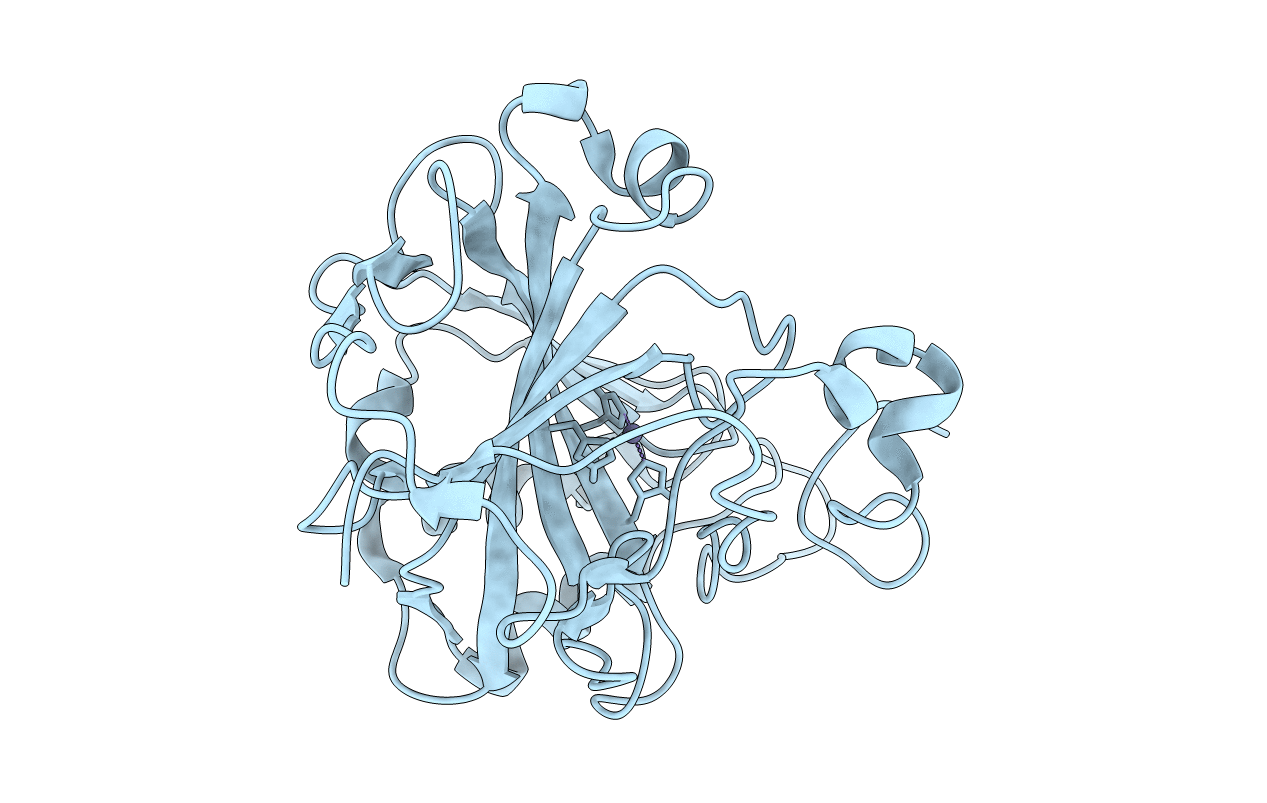
4Y0J image
Deposition Date 2015-02-06
Release Date 2015-04-22
Last Version Date 2024-01-10
Entry Detail
PDB ID:
4Y0J
Keywords:
Title:
H/D exchanged human carbonic anhydrase II pH 6 room temperature neutron crystal structure.
Biological Source:
Source Organism:
Homo sapiens (Taxon ID: 9606)
Method Details:
Experimental Method:
Resolution:
2.00 Å
R-Value Free:
0.29
R-Value Work:
0.26
Space Group:
P 1 21 1
Macromolecular Entities
Polymer Type:polypeptide(L)
Molecule:Carbonic anhydrase 2
Gene (Uniprot):CA2
Chain IDs:A
Chain Length:258
Number of Molecules:1
Biological Source:Homo sapiens
Primary Citation
Joint neutron crystallographic and NMR solution studies of Tyr residue ionization and hydrogen bonding: Implications for enzyme-mediated proton transfer.
Proc.Natl.Acad.Sci.USA 112 5673 5678 (2015)
PMID: 25902526 DOI: 10.1073/pnas.1502255112

Abstact

Human carbonic anhydrase II (HCA II) uses a Zn-bound OH(-)/H2O mechanism to catalyze the reversible hydration of CO2. This catalysis also involves a separate proton transfer step, mediated by an ordered solvent network coordinated by hydrophilic residues. One of these residues, Tyr7, was previously shown to be deprotonated in the neutron crystal structure at pH 10. This observation indicated that Tyr7 has a perturbed pKa compared with free tyrosine. To further probe the pKa of this residue, NMR spectroscopic measurements of [(13)C]Tyr-labeled holo HCA II (with active-site Zn present) were preformed to titrate all Tyr residues between pH 5.4-11.0. In addition, neutron studies of apo HCA II (with Zn removed from the active site) at pH 7.5 and holo HCA II at pH 6 were conducted. This detailed interrogation of tyrosines in HCA II by NMR and neutron crystallography revealed a significantly lowered pKa of Tyr7 and how pH and Tyr proximity to Zn affect hydrogen-bonding interactions.

Legend

Protein

Chemical

Disease

Primary Citation of related structures