4XZ3 image
Deposition Date 2015-02-03
Release Date 2016-01-27
Last Version Date 2024-11-13
Entry Detail
PDB ID:
4XZ3
Keywords:
Title:
Ca. Korarchaeum cryptofilum dinucleotide forming Acetyl-coenzyme A synthetase 1 (Se-Met derivative) in complex with coenzyme A and Mg-AMPPCP, phosphohistidine segment pointing towards nucleotide binding site
Biological Source:
Source Organism:
Method Details:
Experimental Method:
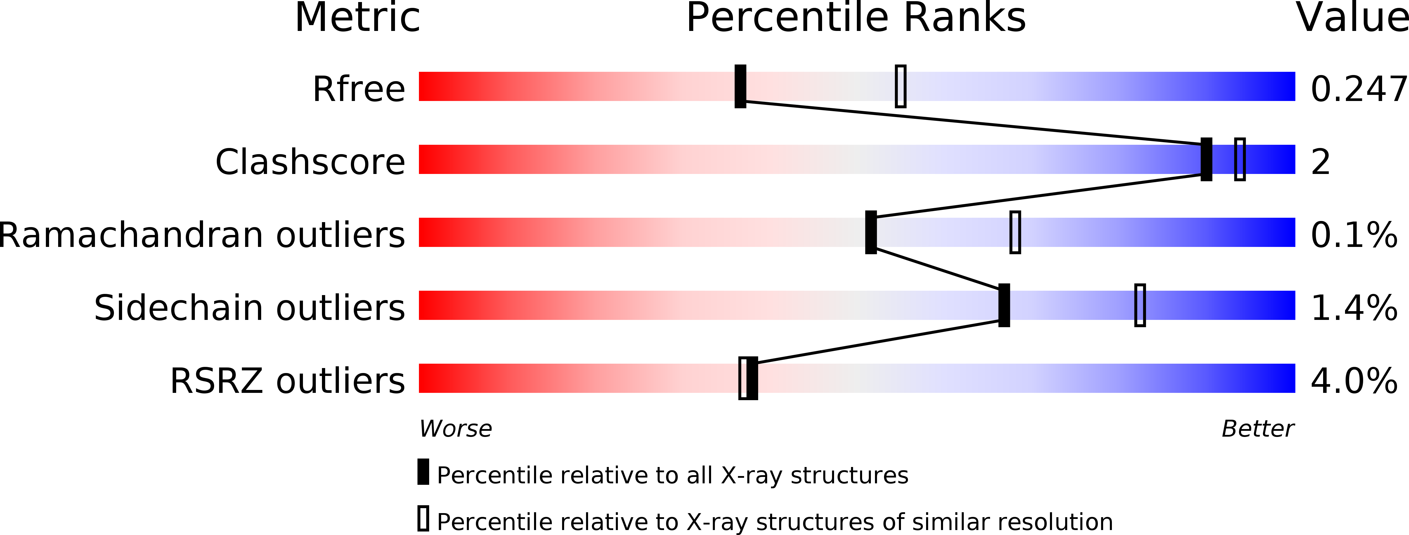
Resolution:
2.40 Å
R-Value Free:
0.24
R-Value Work:
0.19
R-Value Observed:
0.19
Space Group:
P 21 21 21
Macromolecular Entities
Polymer Type:polypeptide(L)
Molecule:Acyl-CoA synthetase (NDP forming)
Gene (Uniprot):Kcr_0198
Chain IDs:A, C
Chain Length:464
Number of Molecules:2
Biological Source:Candidatus Korarchaeum cryptofilum OPF8
Polymer Type:polypeptide(L)
Molecule:Uncharacterized protein
Gene (Uniprot):Kcr_0115
Chain IDs:B, D
Chain Length:230
Number of Molecules:2
Biological Source:Candidatus Korarchaeum cryptofilum OPF8
Modified Residue
Compound ID Chain ID Parent Comp ID Details 2D Image
MSE A MET modified residue
Primary Citation
Structure of NDP-forming Acetyl-CoA synthetase ACD1 reveals a large rearrangement for phosphoryl transfer.
Proc.Natl.Acad.Sci.USA 113 E519 E528 (2016)
PMID: 26787904 DOI: 10.1073/pnas.1518614113

Abstact

The NDP-forming acyl-CoA synthetases (ACDs) catalyze the conversion of various CoA thioesters to the corresponding acids, conserving their chemical energy in form of ATP. The ACDs are the major energy-conserving enzymes in sugar and peptide fermentation of hyperthermophilic archaea. They are considered to be primordial enzymes of ATP synthesis in the early evolution of life. We present the first crystal structures, to our knowledge, of an ACD from the hyperthermophilic archaeon Candidatus Korachaeum cryptofilum. These structures reveal a unique arrangement of the ACD subunits alpha and beta within an α2β2-heterotetrameric complex. This arrangement significantly differs from other members of the superfamily. To transmit an activated phosphoryl moiety from the Ac-CoA binding site (within the alpha subunit) to the NDP-binding site (within the beta subunit), a distance of 51 Å has to be bridged. This transmission requires a larger rearrangement within the protein complex involving a 21-aa-long phosphohistidine-containing segment of the alpha subunit. Spatial restraints of the interaction of this segment with the beta subunit explain the necessity for a second highly conserved His residue within the beta subunit. The data support the proposed four-step reaction mechanism of ACDs, coupling acyl-CoA thioesters with ATP synthesis. Furthermore, the determined crystal structure of the complex with bound Ac-CoA allows first insight, to our knowledge, into the determinants for acyl-CoA substrate specificity. The composition and size of loops protruding into the binding pocket of acyl-CoA are determined by the individual arrangement of the characteristic subdomains.

Legend

Protein

Chemical

Disease

Primary Citation of related structures