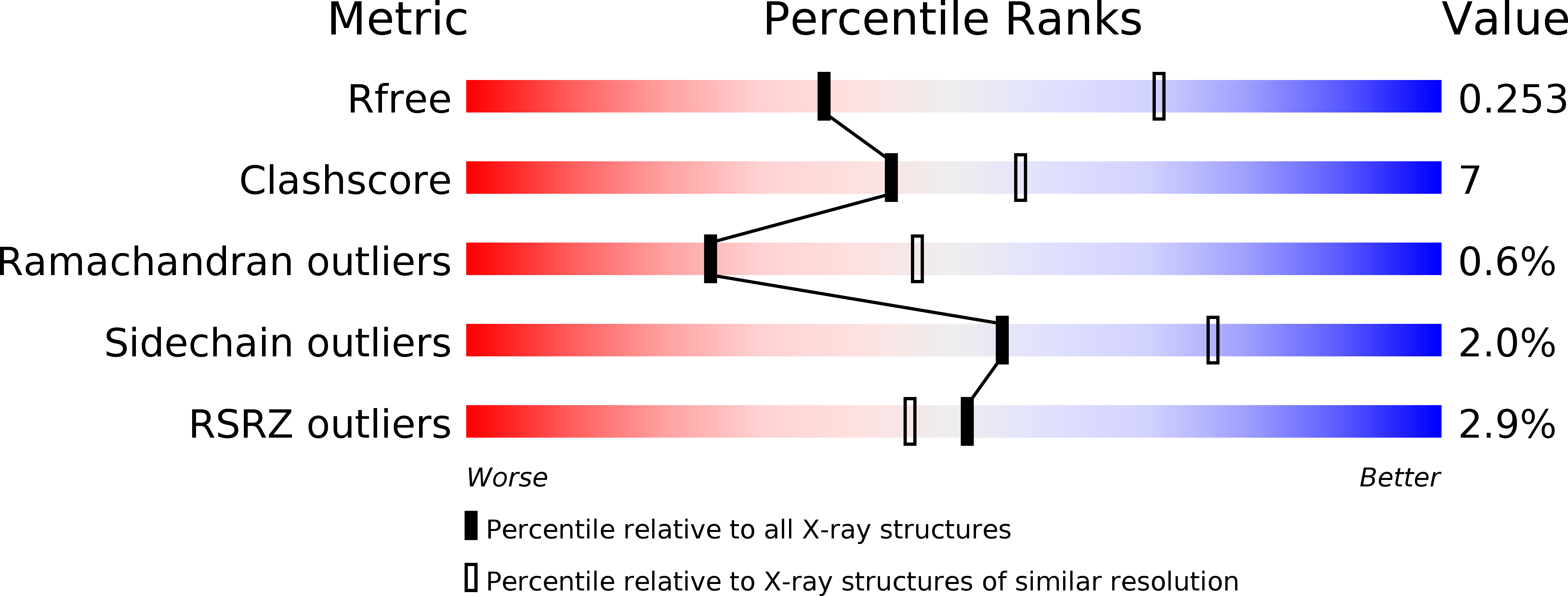
4XXO image
Deposition Date 2015-01-30
Release Date 2015-04-29
Last Version Date 2024-11-06
Entry Detail
PDB ID:
4XXO
Keywords:
Title:
Crystal Structure of Human APOBEC3A
Biological Source:
Source Organism:
Homo sapiens (Taxon ID: 9606)
Host Organism:
Method Details:
Experimental Method:
Resolution:
2.84 Å
R-Value Free:
0.25
R-Value Work:
0.21
R-Value Observed:
0.21
Space Group:
P 65 2 2
Macromolecular Entities
Structures with similar UniProt ID
Protein Blast
Polymer Type:polypeptide(L)
Molecule:DNA dC->dU-editing enzyme APOBEC-3A
Gene (Uniprot):APOBEC3A
Mutagens:E72A, C171A
Chain IDs:A, B
Chain Length:203
Number of Molecules:2
Biological Source:Homo sapiens
Primary Citation
The ssDNA Mutator APOBEC3A Is Regulated by Cooperative Dimerization.
Structure 23 903 911 (2015)
PMID: 25914058 DOI: 10.1016/j.str.2015.03.016

Abstact

Deaminase activity mediated by the human APOBEC3 family of proteins contributes to genomic instability and cancer. APOBEC3A is by far the most active in this family and can cause rapid cell death when overexpressed, but in general how the activity of APOBEC3s is regulated on a molecular level is unclear. In this study, the biochemical and structural basis of APOBEC3A substrate binding and specificity is elucidated. We find that specific binding of single-stranded DNA is regulated by the cooperative dimerization of APOBEC3A. The crystal structure elucidates this homodimer as a symmetric domain swap of the N-terminal residues. This dimer interface provides insights into how cooperative protein-protein interactions may affect function in the APOBEC3 enzymes and provides a potential scaffold for strategies aimed at reducing their mutation load.

Legend

Protein

Chemical

Disease

Primary Citation of related structures