4XXB image
Deposition Date 2015-01-30
Release Date 2015-08-12
Last Version Date 2023-11-08
Entry Detail
PDB ID:
4XXB
Title:
Crystal structure of human MDM2-RPL11
Biological Source:
Source Organism:
Homo sapiens (Taxon ID: 9606)
Host Organism:
Method Details:
Experimental Method:
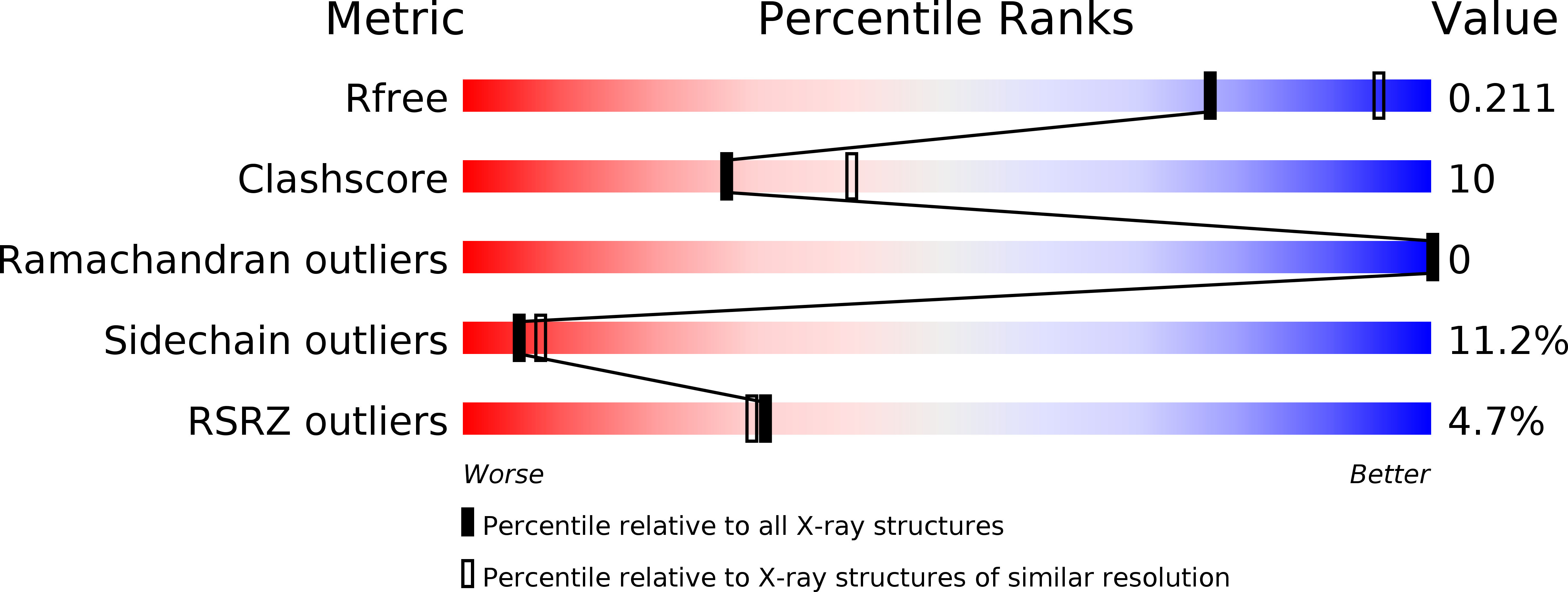
Resolution:
2.40 Å
R-Value Free:
0.22
R-Value Work:
0.19
R-Value Observed:
0.19
Space Group:
P 32 2 1
Macromolecular Entities
Polymer Type:polypeptide(L)
Molecule:60S ribosomal protein L11
Gene (Uniprot):RPL11
Chain IDs:A
Chain Length:178
Number of Molecules:1
Biological Source:Homo sapiens
Polymer Type:polypeptide(L)
Molecule:E3 ubiquitin-protein ligase Mdm2
Gene (Uniprot):MDM2
Chain IDs:B
Chain Length:148
Number of Molecules:1
Biological Source:Homo sapiens
Primary Citation
Structure of human MDM2 complexed with RPL11 reveals the molecular basis of p53 activation
Genes Dev. 29 1524 1534 (2015)
PMID: 26220995 DOI: 10.1101/gad.261792.115

Abstact

The central region of MDM2 is critical for p53 activation and tumor suppression. Upon ribosomal stress, this region is bound by ribosomal proteins, particularly ribosomal protein L11 (RPL11), leading to MDM2 inactivation and subsequent p53 activation. Here, we solved the complex structure of human MDM2-RPL11 at 2.4 Å. MDM2 extensively interacts with RPL11 through an acidic domain and two zinc fingers. Formation of the MDM2-RPL11 complex induces substantial conformational changes in both proteins. RPL11, unable to bind MDM2 mutants, fails to induce the activation of p53 in cells. MDM2 mimics 28S rRNA binding to RPL11. The C4 zinc finger determines RPL11 binding to MDM2 but not its homolog, MDMX. Our results highlight the essential role of the RPL11-MDM2 interaction in p53 activation and tumor suppression and provide a structural basis for potential new anti-tumor drug development.

Legend

Protein

Chemical

Disease

Primary Citation of related structures