
Deposition Date
2015-01-29
Release Date
2015-12-16
Last Version Date
2024-03-20
Entry Detail
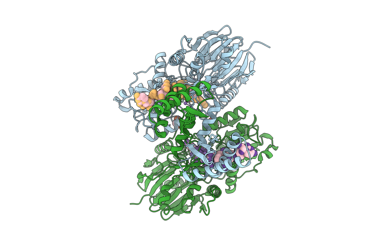
PDB ID:
4XWW
Keywords:
Title:
Crystal structure of RNase J complexed with RNA
Biological Source:
Source Organism(s):
Deinococcus radiodurans (Taxon ID: 1299)
synthetic construct (Taxon ID: 32630)
synthetic construct (Taxon ID: 32630)
Expression System(s):
Method Details:
Experimental Method:
Resolution:
1.70 Å
R-Value Free:
0.21
R-Value Work:
0.19
R-Value Observed:
0.19
Space Group:
P 21 21 21
