4XWM image
Deposition Date 2015-01-29
Release Date 2015-10-28
Last Version Date 2023-11-08
Entry Detail
PDB ID:
4XWM
Keywords:
Title:
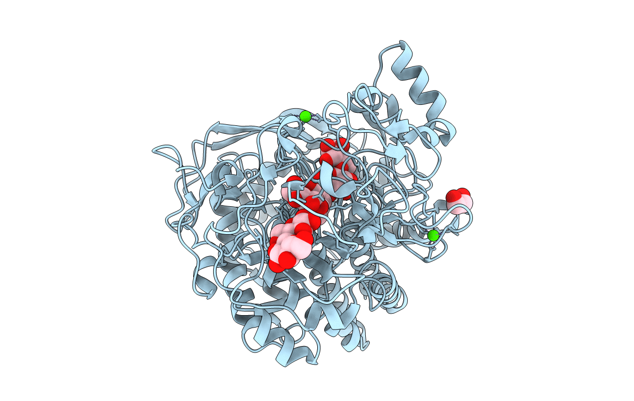
Complex structure of catalytic domain of Clostridium Cellulovorans Exgs and Cellobiose
Biological Source:
Source Organism:
Host Organism:
Method Details:
Experimental Method:
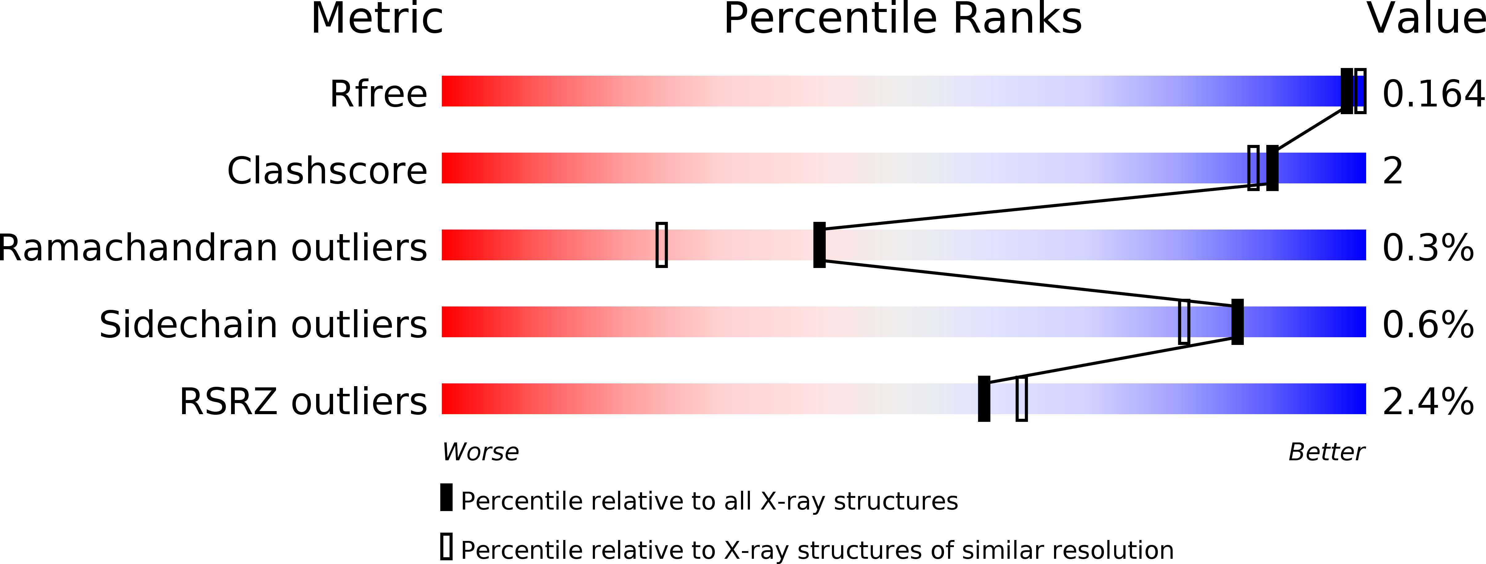
Resolution:
1.70 Å
R-Value Free:
0.16
R-Value Work:
0.14
R-Value Observed:
0.14
Space Group:
P 41 21 2
Macromolecular Entities
Polymer Type:polypeptide(L)
Molecule:Exoglucanase S
Gene (Uniprot):exgS
Chain IDs:A
Chain Length:681
Number of Molecules:1
Biological Source:Clostridium cellulovorans
Peptide-like Molecules
PRD_900005
PRD_900023
Primary Citation
Structures of exoglucanase from Clostridium cellulovorans: cellotetraose binding and cleavage
Acta Crystallogr.,Sect.F 71 1264 1272 (2015)
PMID: 26457517 DOI: 10.1107/S2053230X15015915

Abstact

Exoglucanase/cellobiohydrolase (EC 3.2.1.176) hydrolyzes a β-1,4-glycosidic bond from the reducing end of cellulose and releases cellobiose as the major product. Three complex crystal structures of the glycosyl hydrolase 48 (GH48) cellobiohydrolase S (ExgS) from Clostridium cellulovorans with cellobiose, cellotetraose and triethylene glycol molecules were solved. The product cellobiose occupies subsites +1 and +2 in the open active-site cleft of the enzyme-cellotetraose complex structure, indicating an enzymatic hydrolysis function. Moreover, three triethylene glycol molecules and one pentaethylene glycol molecule are located at active-site subsites -2 to -6 in the structure of the ExgS-triethylene glycol complex shown here. Modelling of glucose into subsite -1 in the active site of the ExgS-cellobiose structure revealed that Glu50 acts as a proton donor and Asp222 plays a nucleophilic role.

Legend

Protein

Chemical

Disease

Primary Citation of related structures