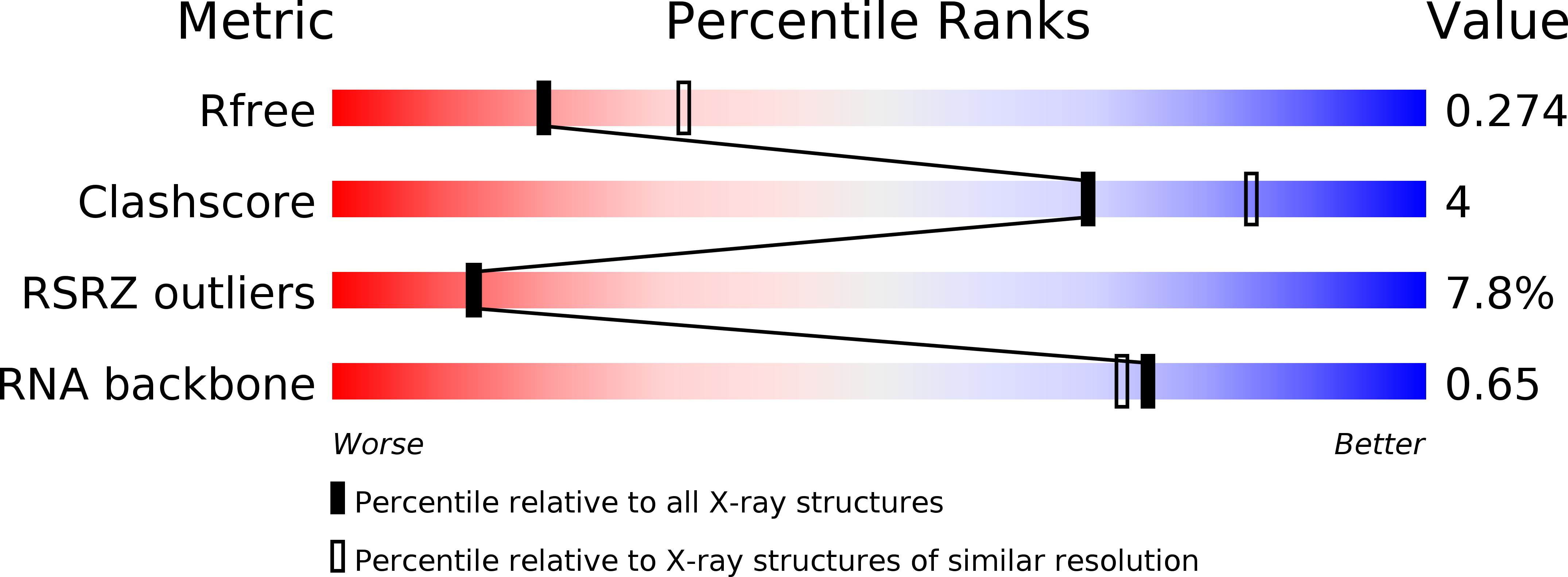
4XW7 image
Entry Detail
PDB ID:
4XW7
Keywords:
Title:
Crystal structure of the ZMP riboswitch at 2.50 angstrom
Biological Source:
Source Organism:
PDB Version:
Deposition Date:
2015-01-28
Release Date:
2015-09-09
Method Details:
Experimental Method:
Resolution:
2.50 Å
R-Value Free:
0.27
R-Value Work:
0.24
R-Value Observed:
0.25
Space Group:
P 43 21 2
Macromolecular Entities
Polymer Type:polyribonucleotide
Description:pfl RNA
Chain IDs:A
Chain Length:64
Number of Molecules:1
Biological Source:Actinomyces odontolyticus
Primary Citation
Metal Ion-Mediated Nucleobase Recognition by the ZTP Riboswitch.
Chem.Biol. 22 829 837 (2015)
PMID: 26144884 DOI: 10.1016/j.chembiol.2015.06.007

Abstact

The ZTP riboswitch is a widespread family of regulatory RNAs that upregulate de novo purine synthesis in response to increased intracellular levels of ZTP or ZMP. As an important intermediate in purine biosynthesis, ZMP also serves as a proxy for the concentration of N10-formyl-tetrahydrofolate, a key component of one-carbon metabolism. Here, we report the structure of the ZTP riboswitch bound to ZMP at a resolution of 1.80 Å. The RNA contains two subdomains brought together through a long-range pseudoknot further stabilized through helix-helix packing. ZMP is bound at the subdomain interface of the RNA through a set of interactions with the base, ribose sugar, and phosphate moieties of the ligand. Unique to nucleobase recognition by RNAs, the Z base is inner-sphere coordinated to a magnesium cation bound by two backbone phosphates. This interaction, along with steric hindrance by the backbone, imparts specificity over chemically similar compounds such as ATP/AMP.

Legend

Protein

Chemical

Disease

Primary Citation of related structures