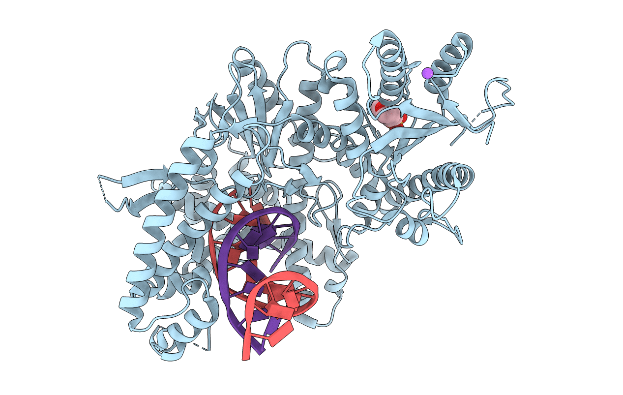
4XVL image
Entry Detail
PDB ID:
4XVL
Keywords:
Title:
Binary complex of human polymerase nu and DNA with the finger domain open
Biological Source:
Source Organism:
Host Organism:
PDB Version:
Deposition Date:
2015-01-27
Release Date:
2015-03-11
Method Details:
Experimental Method:
Resolution:
3.30 Å
R-Value Free:
0.22
R-Value Work:
0.20
R-Value Observed:
0.20
Space Group:
H 3 2
Macromolecular Entities
Polymer Type:polypeptide(L)
Description:DNA polymerase nu
Chain IDs:A
Chain Length:666
Number of Molecules:1
Biological Source:Homo sapiens
Polymer Type:polydeoxyribonucleotide
Description:DNA (5'-D(*GP*AP*TP*CP*TP*GP*AP*CP*GP*CP*TP*AP*CP*G)-3')
Chain IDs:B (auth: P)
Chain Length:14
Number of Molecules:1
Biological Source:Homo sapiens
Polymer Type:polydeoxyribonucleotide
Description:DNA (5'-D(*TP*CP*GP*TP*AP*GP*CP*GP*TP*CP*A)-3')
Chain IDs:C (auth: T)
Chain Length:10
Number of Molecules:1
Biological Source:Homo sapiens
Primary Citation
How a homolog of high-fidelity replicases conducts mutagenic DNA synthesis.
Nat.Struct.Mol.Biol. 22 298 303 (2015)
PMID: 25775266 DOI: 10.1038/nsmb.2985

Abstact

All DNA replicases achieve high fidelity by a conserved mechanism, but each translesion polymerase carries out mutagenic DNA synthesis in its own way. Here we report crystal structures of human DNA polymerase ν (Pol ν), which is homologous to high-fidelity replicases yet is error prone. Instead of a simple open-to-closed movement of the O helix upon binding of a correct incoming nucleotide, Pol ν has a different open state and requires the finger domain to swing sideways and undergo both opening and closing motions to accommodate the nascent base pair. A single-amino acid substitution in the O helix of the finger domain improves the fidelity of Pol ν nearly ten-fold. A unique cavity and the flexibility of the thumb domain allow Pol ν to generate and accommodate a looped-out primer strand. Primer loop-out may be a mechanism for DNA trinucloetide-repeat expansion.

Legend

Protein

Chemical

Disease

Primary Citation of related structures