4XUF image
Deposition Date 2015-01-25
Release Date 2015-04-15
Last Version Date 2023-09-27
Entry Detail
PDB ID:
4XUF
Title:
Crystal structure of the FLT3 kinase domain bound to the inhibitor quizartinib (AC220)
Biological Source:
Source Organism:
Homo sapiens (Taxon ID: 9606)
Method Details:
Experimental Method:
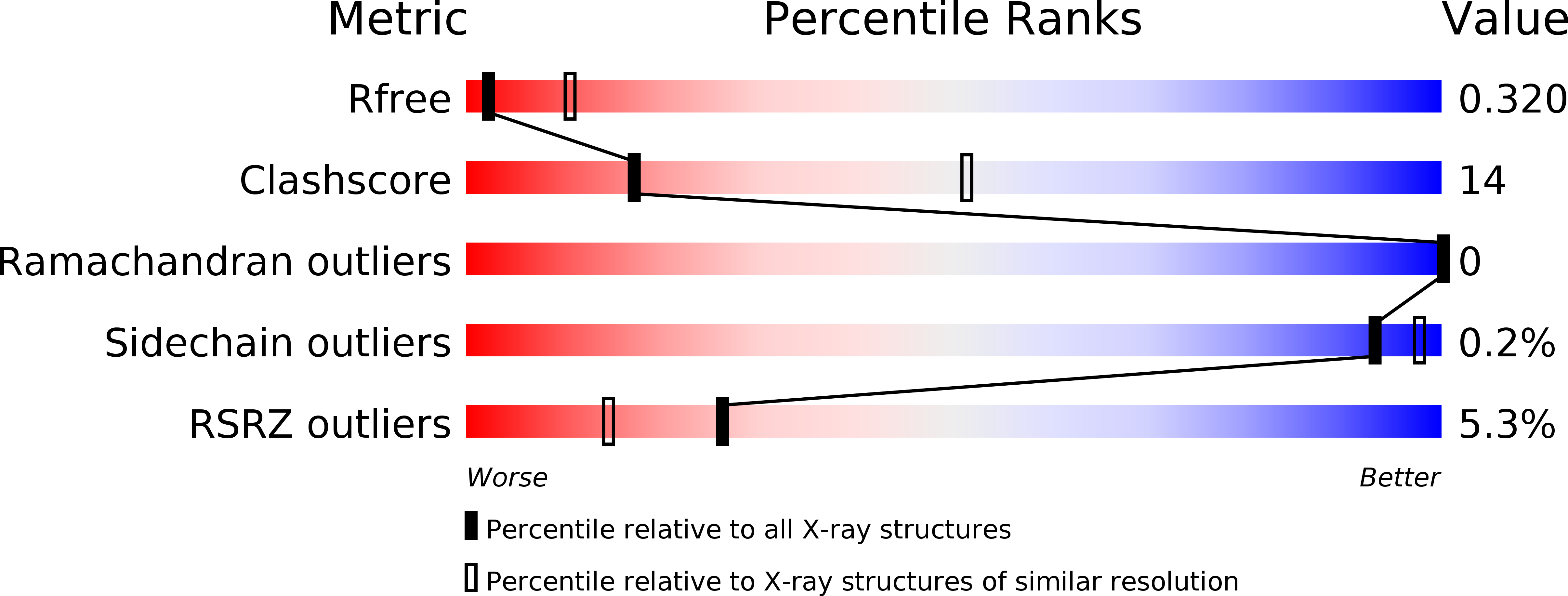
Resolution:
3.20 Å
R-Value Free:
0.32
R-Value Work:
0.29
R-Value Observed:
0.29
Space Group:
P 21 21 21
Macromolecular Entities
Structures with similar UniProt ID
Protein Blast
Polymer Type:polypeptide(L)
Molecule:Receptor-type tyrosine-protein kinase FLT3
Gene (Uniprot):FLT3
Mutagens:delta(711-761)
Chain IDs:A, B
Chain Length:297
Number of Molecules:2
Biological Source:Homo sapiens
Ligand Molecules
Primary Citation
Crystal Structure of the FLT3 Kinase Domain Bound to the Inhibitor Quizartinib (AC220).
Plos One 10 e0121177 e0121177 (2015)
PMID: 25837374 DOI: 10.1371/journal.pone.0121177

Abstact

More than 30% of acute myeloid leukemia (AML) patients possess activating mutations in the receptor tyrosine kinase FMS-like tyrosine kinase 3 or FLT3. A small-molecule inhibitor of FLT3 (known as quizartinib or AC220) that is currently in clinical trials appears promising for the treatment of AML. Here, we report the co-crystal structure of the kinase domain of FLT3 in complex with quizartinib. FLT3 with quizartinib bound adopts an "Abl-like" inactive conformation with the activation loop stabilized in the "DFG-out" orientation and folded back onto the kinase domain. This conformation is similar to that observed for the uncomplexed intracellular domain of FLT3 as well as for related receptor tyrosine kinases, except for a localized induced fit in the activation loop. The co-crystal structure reveals the interactions between quizartinib and the active site of FLT3 that are key for achieving its high potency against both wild-type FLT3 as well as a FLT3 variant observed in many AML patients. This co-complex further provides a structural rationale for quizartinib-resistance mutations.

Legend

Protein

Chemical

Disease

Primary Citation of related structures
Feedback Form
Name
Email
Institute
Feedback