Deposition Date
2015-01-25
Release Date
2015-10-14
Last Version Date
2024-03-20
Entry Detail
PDB ID:
4XU5
Keywords:
Title:
Crystal structure of MvINS bound to a bromine-derived 14C Diacylglycerol (DAG) at 2.1A resolution
Biological Source:
Source Organism(s):
Mycobacterium vanbaalenii PYR-1 (Taxon ID: 350058)
Expression System(s):
Method Details:
Experimental Method:
Resolution:
2.10 Å
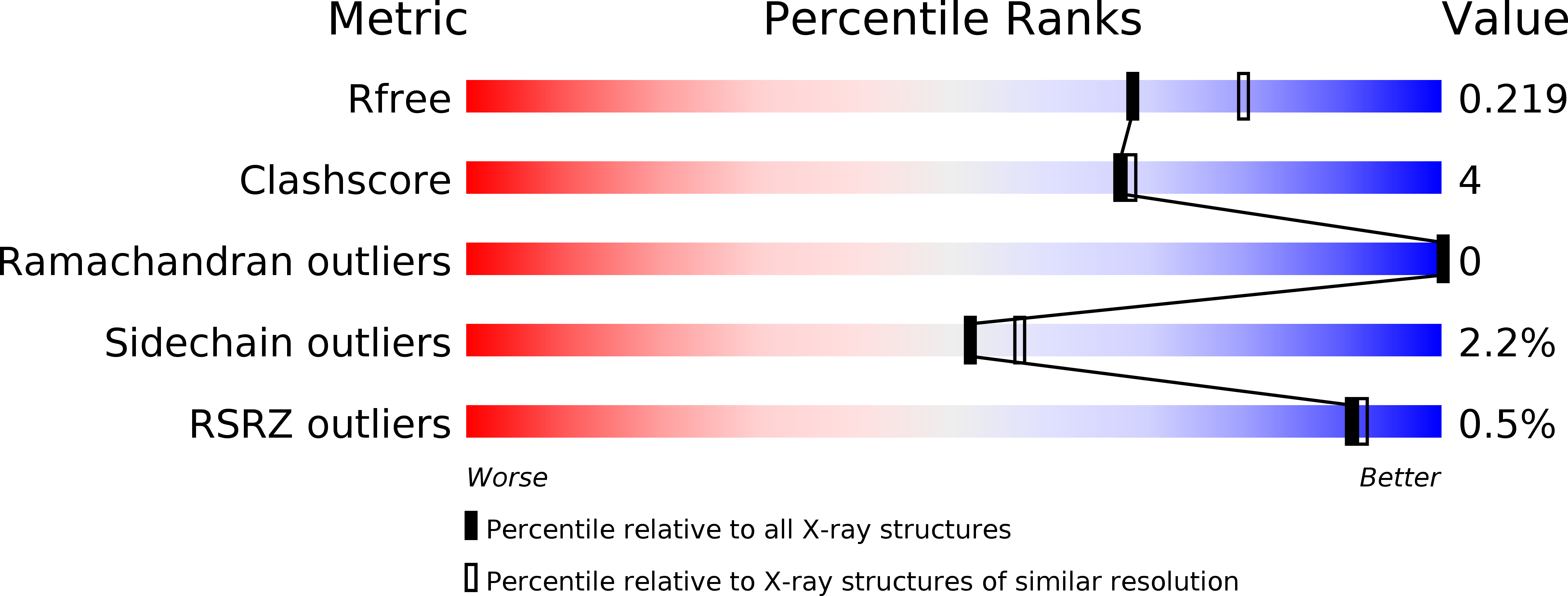
R-Value Free:
0.22
R-Value Work:
0.19
R-Value Observed:
0.20
Space Group:
H 3