4XTQ image
Entry Detail
PDB ID:
4XTQ
Title:
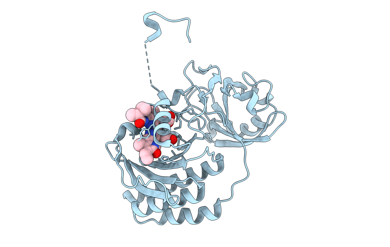
Crystal structure of a mutant (C20S) of a near-infrared fluorescent protein BphP1-FP
Biological Source:
Host Organism:
PDB Version:
Deposition Date:
2015-01-23
Release Date:
2015-12-09
Method Details:
Experimental Method:
Resolution:
1.64 Å
R-Value Free:
0.20
R-Value Work:
0.17
R-Value Observed:
0.17
Space Group:
P 21 21 21
Macromolecular Entities
Polymer Type:polypeptide(L)
Description:BphP1-FP/C20S
Mutations:C20S
Chain IDs:A
Chain Length:327
Number of Molecules:1
Biological Source:Rhodopseudomonas palustris
Primary Citation
Molecular Basis of Spectral Diversity in Near-Infrared Phytochrome-Based Fluorescent Proteins.
Chem.Biol. 22 1540 1551 (2015)
PMID: 26590639 DOI: 10.1016/j.chembiol.2015.10.007

Abstact

Near-infrared fluorescent proteins (NIR FPs) engineered from bacterial phytochromes (BphPs) are the probes of choice for deep-tissue imaging. Detection of several processes requires spectrally distinct NIR FPs. We developed an NIR FP, BphP1-FP, which has the most blue-shifted spectra and the highest fluorescence quantum yield among BphP-derived FPs. We found that these properties result from the binding of the biliverdin chromophore to a cysteine residue in the GAF domain, unlike natural BphPs and other BphP-based FPs. To elucidate the molecular basis of the spectral shift, we applied biochemical, structural and mass spectrometry analyses and revealed the formation of unique chromophore species. Mutagenesis of NIR FPs of different origins indicated that the mechanism of the spectral shift is general and can be used to design multicolor NIR FPs from other BphPs. We applied pairs of spectrally distinct point cysteine mutants to multicolor cell labeling and demonstrated that they perform well in model deep-tissue imaging.

Legend

Protein

Chemical

Disease

Primary Citation of related structures