4XQM image
Deposition Date 2015-01-19
Release Date 2015-07-22
Last Version Date 2024-10-09
Entry Detail
PDB ID:
4XQM
Keywords:
Title:
Crystal structure of the MRH domain of Glucosidase II beta bound to mannose
Biological Source:
Method Details:
Experimental Method:
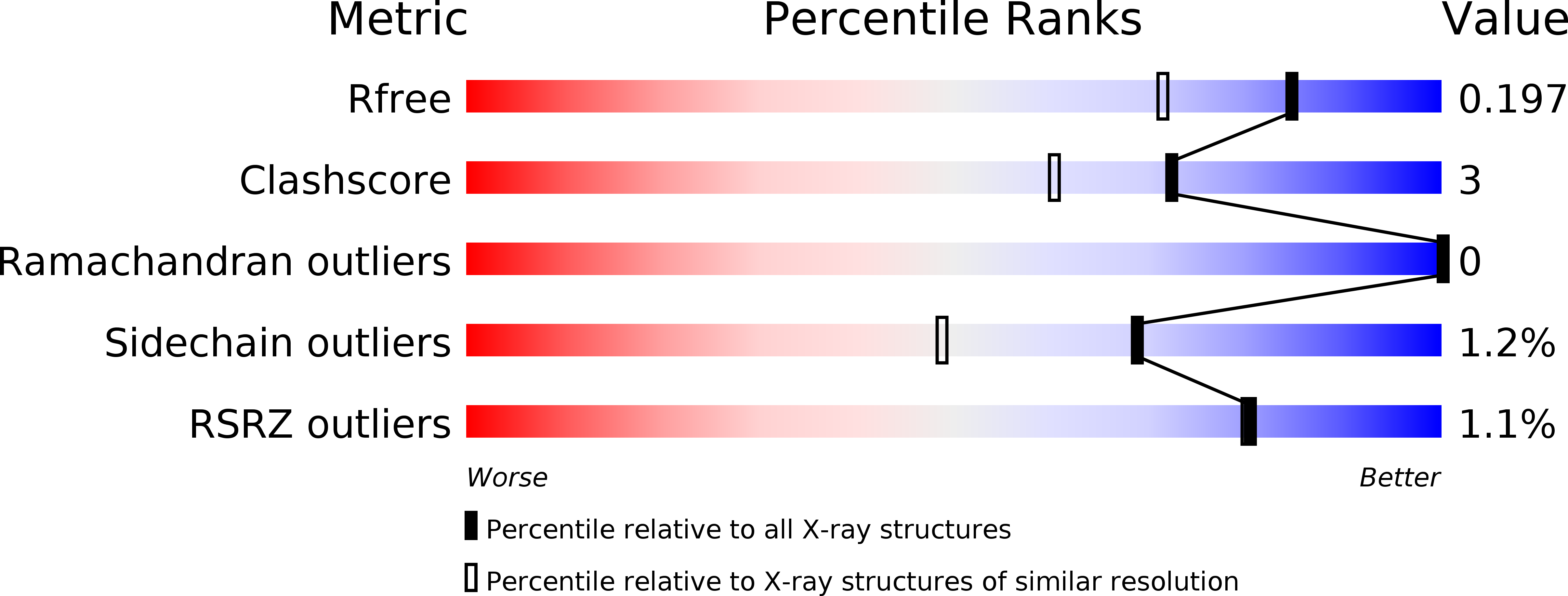
Resolution:
1.63 Å
R-Value Free:
0.19
R-Value Work:
0.16
R-Value Observed:
0.17
Space Group:
I 4
Macromolecular Entities
Structures with similar UniProt ID
Protein Blast
Polymer Type:polypeptide(L)
Molecule:Glucosidase 2 subunit beta
Gene (Uniprot):gtb1
Chain IDs:A
Chain Length:94
Number of Molecules:1
Biological Source:Schizosaccharomyces pombe (strain 972 / ATCC 24843)
Ligand Molecules
Primary Citation
Crystal Structure and Functional Analyses of the Lectin Domain of Glucosidase II: Insights into Oligomannose Recognition.
Biochemistry 54 4097 4111 (2015)
PMID: 26062005 DOI: 10.1021/acs.biochem.5b00256

Abstact

N-Glycans are modified as part of a quality control mechanism during glycoprotein folding in the endoplasmic reticulum (ER). Glucosidase II (GII) plays a critical role by generating monoglucosylated glycans that are recognized by lectin chaperones, calnexin and calreticulin. To understand how the hydrolytic activity of GIIα is enhanced by the mannose 6-phosphate receptor (MPR) homology domain (MRH domain) of its β subunit, we now report a 1.6 Å resolution crystal structure of the MRH domain of GIIβ bound to mannose. A comparison of ligand-bound and unbound structures reveals no major difference in their overall fold, but rather a repositioning of side chains throughout the binding pocket, including Y372. Mutation of Y372 inhibits GII activity, demonstrating an important role for Y372 in regulating GII activity. Comparison of the MRH domains of GIIβ, MPRs, and the ER lectin OS-9 identified conserved residues that are critical for the structural integrity and architecture of the carbohydrate binding pocket. As shown by nuclear magnetic resonance spectroscopy, mutations of the primary binding pocket residues and adjacent W409, all of which inhibit the activity of GII both in vitro and in vivo, do not cause a significant change in the overall fold of the GIIβ MRH domain but impact locally the stability of the binding pocket. W409 does not directly contact mannose; rather, its indole ring is stabilized by binding into a hydrophobic pocket of an adjacent crystallographic neighbor. This suggests that W409 interacts with a hydrophobic region of the GIIβ or GIIα subunit to modulate its effect on GII activity.

Legend

Protein

Chemical

Disease

Primary Citation of related structures
Feedback Form
Name
Email
Institute
Feedback