Deposition Date
2015-01-16
Release Date
2015-07-29
Last Version Date
2024-10-30
Entry Detail
PDB ID:
4XO1
Keywords:
Title:
crystal structure of Se-Met GnsA with double mutations
Biological Source:
Source Organism:
Escherichia coli (Taxon ID: 83333)
Host Organism:
Method Details:
Experimental Method:
Resolution:
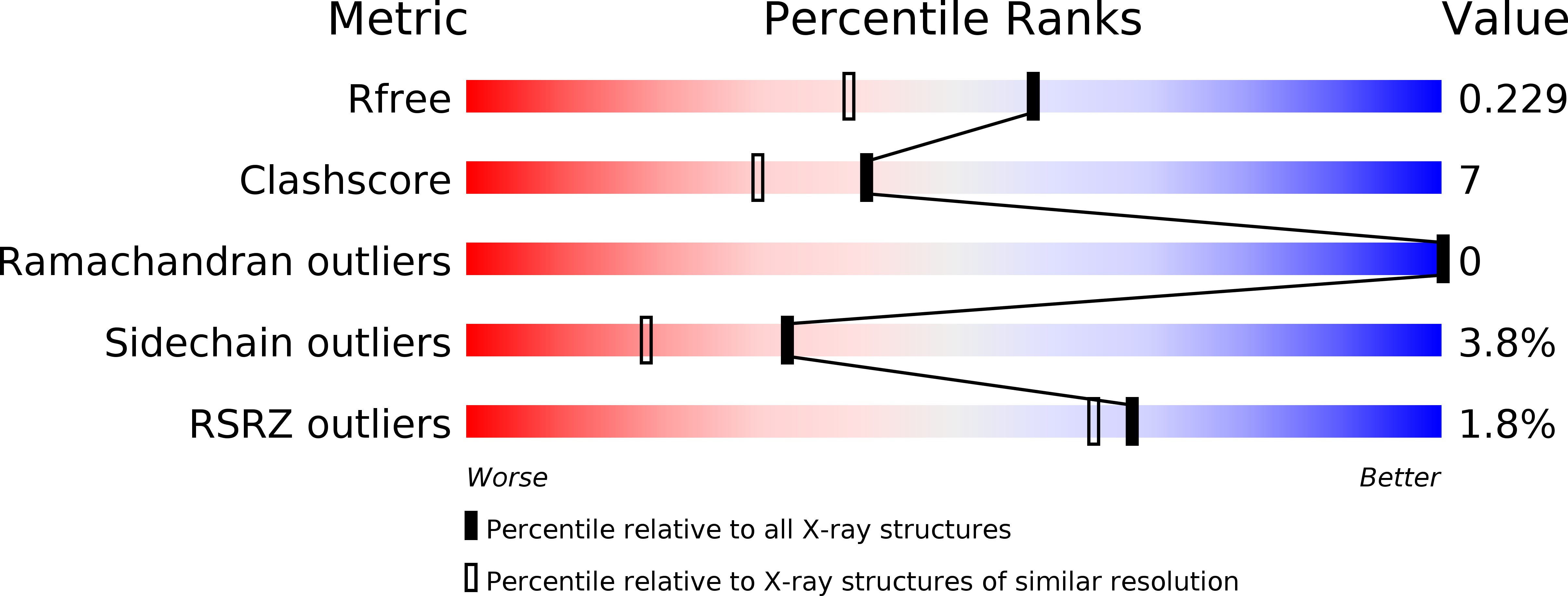
1.80 Å
R-Value Free:
0.22
R-Value Work:
0.19
R-Value Observed:
0.19
Space Group:
C 2 2 21