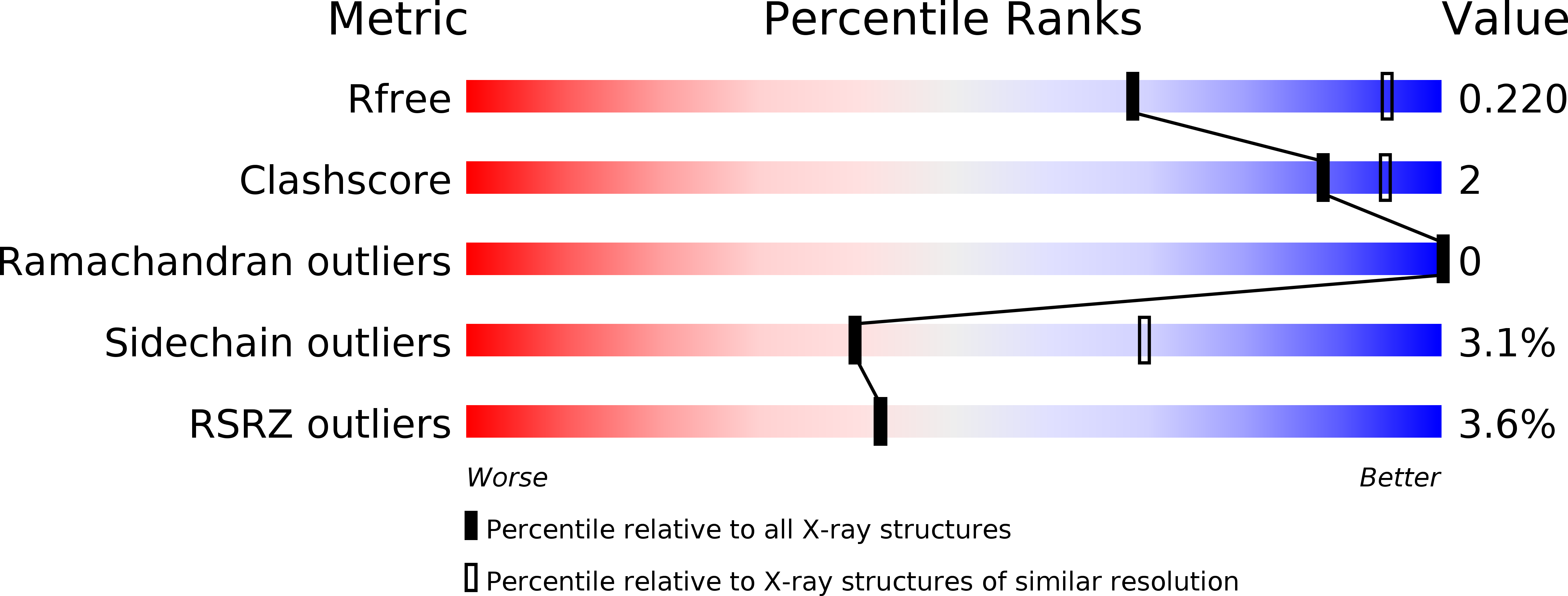
4XNW image
Deposition Date 2015-01-16
Release Date 2015-04-01
Last Version Date 2024-10-23
Entry Detail
PDB ID:
4XNW
Title:
The human P2Y1 receptor in complex with MRS2500
Biological Source:
Source Organism:
Host Organism:
Method Details:
Experimental Method:
Resolution:
2.70 Å
R-Value Free:
0.26
R-Value Work:
0.21
R-Value Observed:
0.22
Space Group:
P 1
Macromolecular Entities
Polymer Type:polypeptide(L)
Molecule:P2Y purinoceptor 1,Rubredoxin,P2Y purinoceptor 1
Gene (Uniprot):P2RY1
Mutations:D320N
Chain IDs:A, B (auth: C)
Chain Length:421
Number of Molecules:2
Biological Source:Homo sapiens, Clostridium pasteurianum
Primary Citation
Two disparate ligand-binding sites in the human P2Y1 receptor
Nature 520 317 321 (2015)
PMID: 25822790 DOI: 10.1038/nature14287

Abstact

In response to adenosine 5'-diphosphate, the P2Y1 receptor (P2Y1R) facilitates platelet aggregation, and thus serves as an important antithrombotic drug target. Here we report the crystal structures of the human P2Y1R in complex with a nucleotide antagonist MRS2500 at 2.7 Å resolution, and with a non-nucleotide antagonist BPTU at 2.2 Å resolution. The structures reveal two distinct ligand-binding sites, providing atomic details of P2Y1R's unique ligand-binding modes. MRS2500 recognizes a binding site within the seven transmembrane bundle of P2Y1R, which is different in shape and location from the nucleotide binding site in the previously determined structure of P2Y12R, representative of another P2YR subfamily. BPTU binds to an allosteric pocket on the external receptor interface with the lipid bilayer, making it the first structurally characterized selective G-protein-coupled receptor (GPCR) ligand located entirely outside of the helical bundle. These high-resolution insights into P2Y1R should enable discovery of new orthosteric and allosteric antithrombotic drugs with reduced adverse effects.

Legend

Protein

Chemical

Disease

Primary Citation of related structures