4XNU image
Entry Detail
PDB ID:
4XNU
Title:
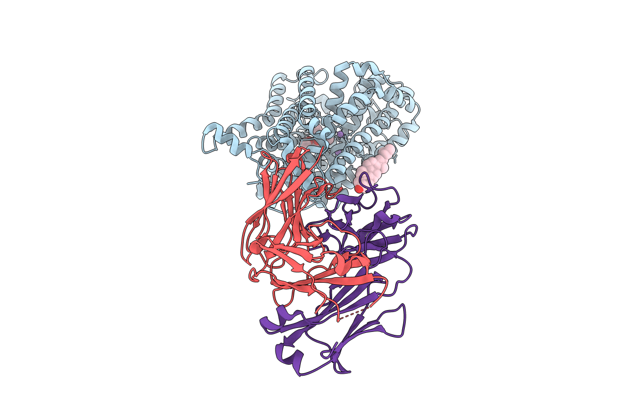
X-ray structure of Drosophila dopamine transporter in complex with nisoxetine
Biological Source:
Host Organism:
PDB Version:
Deposition Date:
2015-01-16
Release Date:
2015-05-13
Method Details:
Experimental Method:
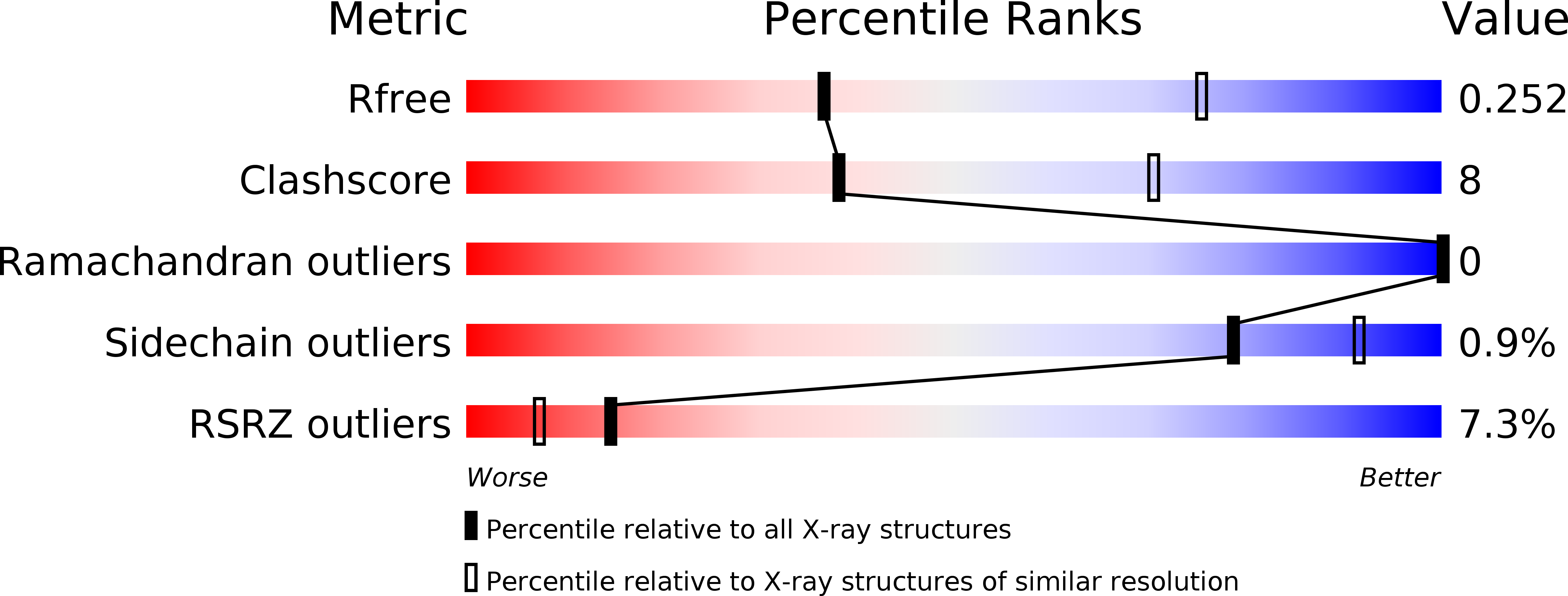
Resolution:
2.98 Å
R-Value Free:
0.25
R-Value Work:
0.22
R-Value Observed:
0.22
Space Group:
P 21 21 21
Macromolecular Entities
Polymer Type:polypeptide(L)
Description:Integral membrane protein-dopamine transporter
Chain IDs:A
Chain Length:532
Number of Molecules:1
Biological Source:Drosophila melanogaster
Polymer Type:polypeptide(L)
Description:antibody fragment heavy chain
Chain IDs:C (auth: H)
Chain Length:240
Number of Molecules:1
Biological Source:Mus musculus
Polymer Type:polypeptide(L)
Description:antibody fragment light chain
Chain IDs:B (auth: L)
Chain Length:214
Number of Molecules:1
Biological Source:Mus musculus
Primary Citation
X-ray structures of Drosophila dopamine transporter in complex with nisoxetine and reboxetine.
Nat.Struct.Mol.Biol. 22 506 508 (2015)
PMID: 25961798 DOI: 10.1038/nsmb.3029

Abstact

Most antidepressants elicit their therapeutic benefits through selective blockade of Na(+)/Cl(-)-coupled neurotransmitter transporters. Here we report X-ray structures of the Drosophila melanogaster dopamine transporter in complexes with the polycyclic antidepressants nisoxetine or reboxetine. The inhibitors stabilize the transporter in an outward-open conformation by occupying the substrate-binding site. These structures explain how interactions between the binding pocket and substituents on the aromatic rings of antidepressants modulate drug-transporter selectivity.

Legend

Protein

Chemical

Disease

Primary Citation of related structures