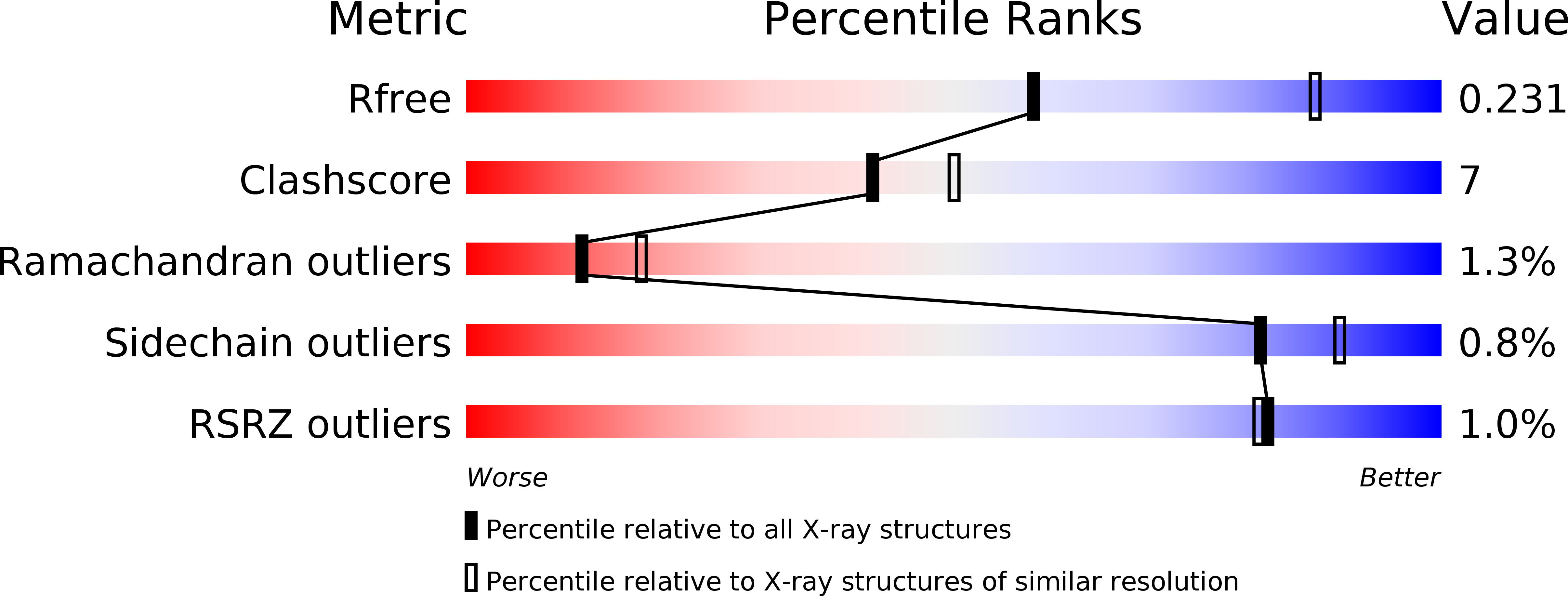
4XGK image
Deposition Date 2014-12-31
Release Date 2015-08-12
Last Version Date 2023-09-27
Entry Detail
PDB ID:
4XGK
Title:
Crystal structure of UDP-galactopyranose mutase from Corynebacterium diphtheriae in complex with 2-[4-(4-chlorophenyl)-7-(2-thienyl)-2-thia-5,6,8,9-tetrazabicyclo[4.3.0]nona-4,7,9-trien-3-yl]acetic
Biological Source:
Source Organism:
Host Organism:
Method Details:
Experimental Method:
Resolution:
2.65 Å
R-Value Free:
0.22
R-Value Work:
0.18
R-Value Observed:
0.18
Space Group:
P 64 2 2
Macromolecular Entities
Polymer Type:polypeptide(L)
Molecule:UDP-galactopyranose mutase
Gene (Uniprot):glf
Chain IDs:A, B
Chain Length:388
Number of Molecules:2
Biological Source:Corynebacterium diphtheriae
Primary Citation
Virtual Screening for UDP-Galactopyranose Mutase Ligands Identifies a New Class of Antimycobacterial Agents.
Acs Chem.Biol. 10 2209 2218 (2015)
PMID: 26214585 DOI: 10.1021/acschembio.5b00370

Abstact

Galactofuranose (Galf) is present in glycans critical for the virulence and viability of several pathogenic microbes, including Mycobacterium tuberculosis, yet the monosaccharide is absent from mammalian glycans. Uridine 5'-diphosphate-galactopyranose mutase (UGM) catalyzes the formation of UDP-Galf, which is required to produce Galf-containing glycoconjugates. Inhibitors of UGM have therefore been sought, both as antimicrobial leads and as tools to delineate the roles of Galf in cells. Obtaining cell permeable UGM probes by either design or high throughput screens has been difficult, as has elucidating how UGM binds small molecule, noncarbohydrate inhibitors. To address these issues, we employed structure-based virtual screening to uncover new inhibitor chemotypes, including a triazolothiadiazine series. These compounds are among the most potent antimycobacterial UGM inhibitors described. They also facilitated determination of a UGM-small molecule inhibitor structure, which can guide optimization. A comparison of results from the computational screen and a high-throughput fluorescence polarization (FP) screen indicated that the scaffold hits from the former had been evaluated in the FP screen but missed. By focusing on promising compounds, the virtual screen rescued false negatives, providing a blueprint for generating new UGM probes and therapeutic leads.

Legend

Protein

Chemical

Disease

Primary Citation of related structures