4XE5 image
Deposition Date 2014-12-22
Release Date 2016-03-09
Last Version Date 2024-11-06
Entry Detail
PDB ID:
4XE5
Keywords:
Title:
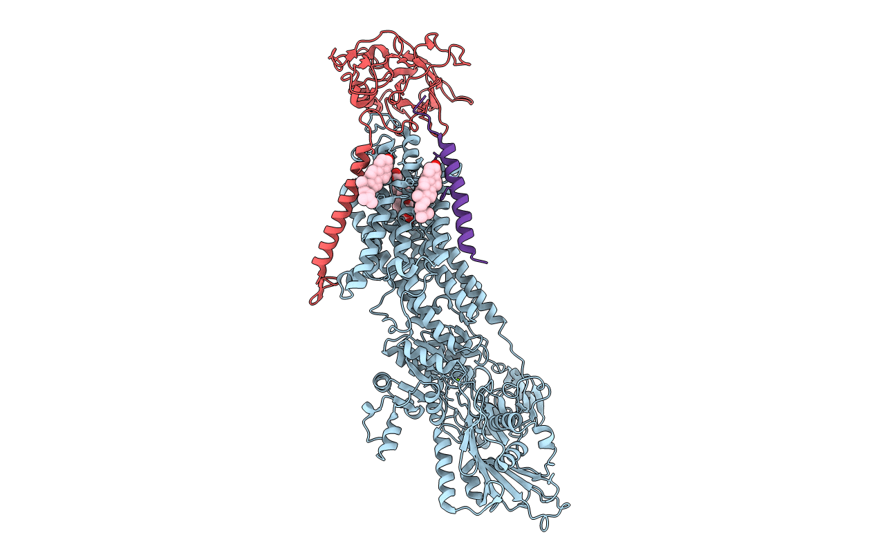
Crystal structure of the Na,K-ATPase from bovine
Biological Source:
Source Organism:
Bos taurus (Taxon ID: 9913)
Method Details:
Experimental Method:
Resolution:
3.90 Å
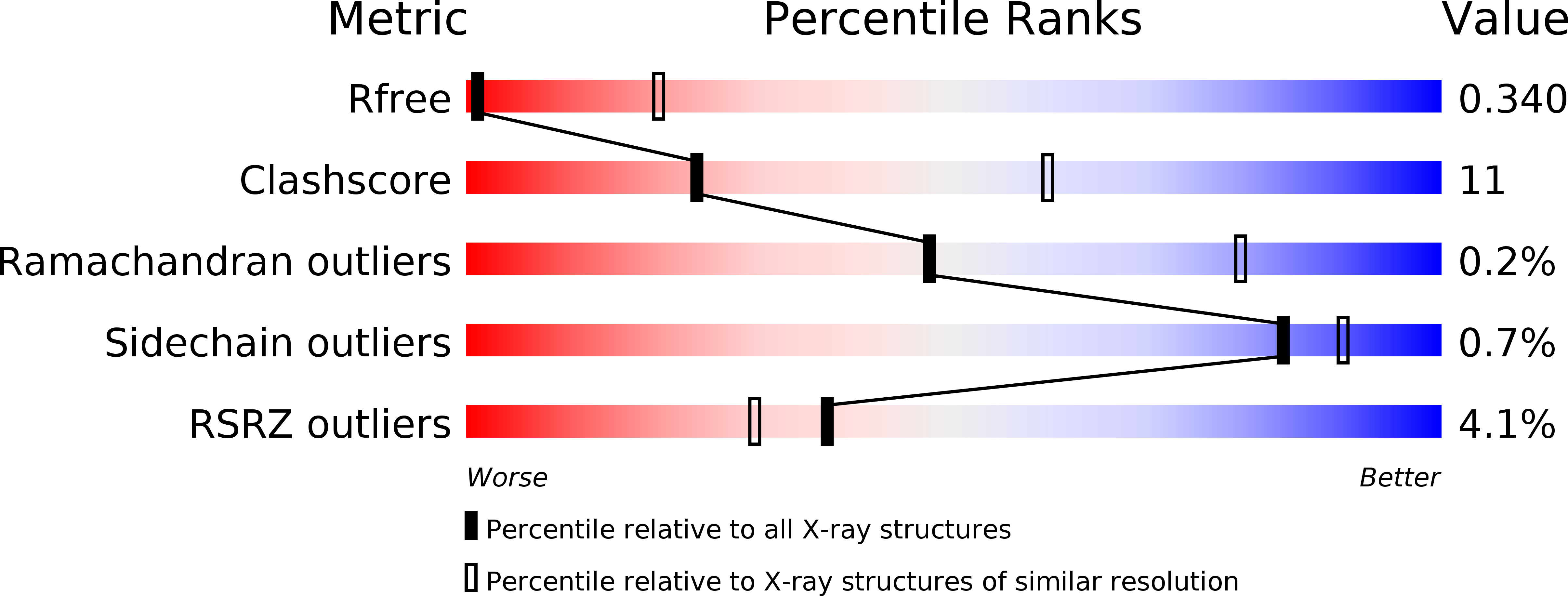
R-Value Free:
0.33
R-Value Work:
0.32
R-Value Observed:
0.32
Space Group:
C 2 2 21
Macromolecular Entities
Structures with similar UniProt ID
Protein Blast
Polymer Type:polypeptide(L)
Molecule:Sodium/potassium-transporting ATPase subunit alpha-1
Gene (Uniprot):ATP1A1
Chain IDs:A
Chain Length:1021
Number of Molecules:1
Biological Source:Bos taurus
Structures with similar UniProt ID
Protein Blast
Polymer Type:polypeptide(L)
Molecule:Sodium/potassium-transporting ATPase subunit beta
Gene (Uniprot):ATP1B1
Chain IDs:B
Chain Length:303
Number of Molecules:1
Biological Source:Bos taurus
Structures with similar UniProt ID
Protein Blast
Polymer Type:polypeptide(L)
Molecule:Sodium/potassium-transporting ATPase subunit gamma
Gene (Uniprot):FXYD2
Chain IDs:C (auth: G)
Chain Length:58
Number of Molecules:1
Biological Source:Bos taurus
Modified Residue
Compound ID Chain ID Parent Comp ID Details 2D Image
BFD A ASP modified residue
Primary Citation
Isolation, crystallization and crystal structure determination of bovine kidney Na(+),K(+)-ATPase.
Acta Crystallogr.,Sect.F 72 282 287 (2016)
PMID: 27050261 DOI: 10.1107/S2053230X1600279X

Abstact

Na(+),K(+)-ATPase is responsible for the transport of Na(+) and K(+) across the plasma membrane in animal cells, thereby sustaining vital electrochemical gradients that energize channels and secondary transporters. The crystal structure of Na(+),K(+)-ATPase has previously been elucidated using the enzyme from native sources such as porcine kidney and shark rectal gland. Here, the isolation, crystallization and first structure determination of bovine kidney Na(+),K(+)-ATPase in a high-affinity E2-BeF3(-)-ouabain complex with bound magnesium are described. Crystals belonging to the orthorhombic space group C2221 with one molecule in the asymmetric unit exhibited anisotropic diffraction to a resolution of 3.7 Å with full completeness to a resolution of 4.2 Å. The structure was determined by molecular replacement, revealing unbiased electron-density features for bound BeF3(-), ouabain and Mg(2+) ions.

Legend

Protein

Chemical

Disease

Primary Citation of related structures