4X4C image
Deposition Date 2014-12-02
Release Date 2015-03-11
Last Version Date 2024-01-10
Entry Detail
PDB ID:
4X4C
Keywords:
Title:
RADIATION DAMAGE TO THE NUCLEOPROTEIN COMPLEX C.Esp1396I: DOSE (DWD) 6.2 MGy
Biological Source:
Source Organism:
Method Details:
Experimental Method:
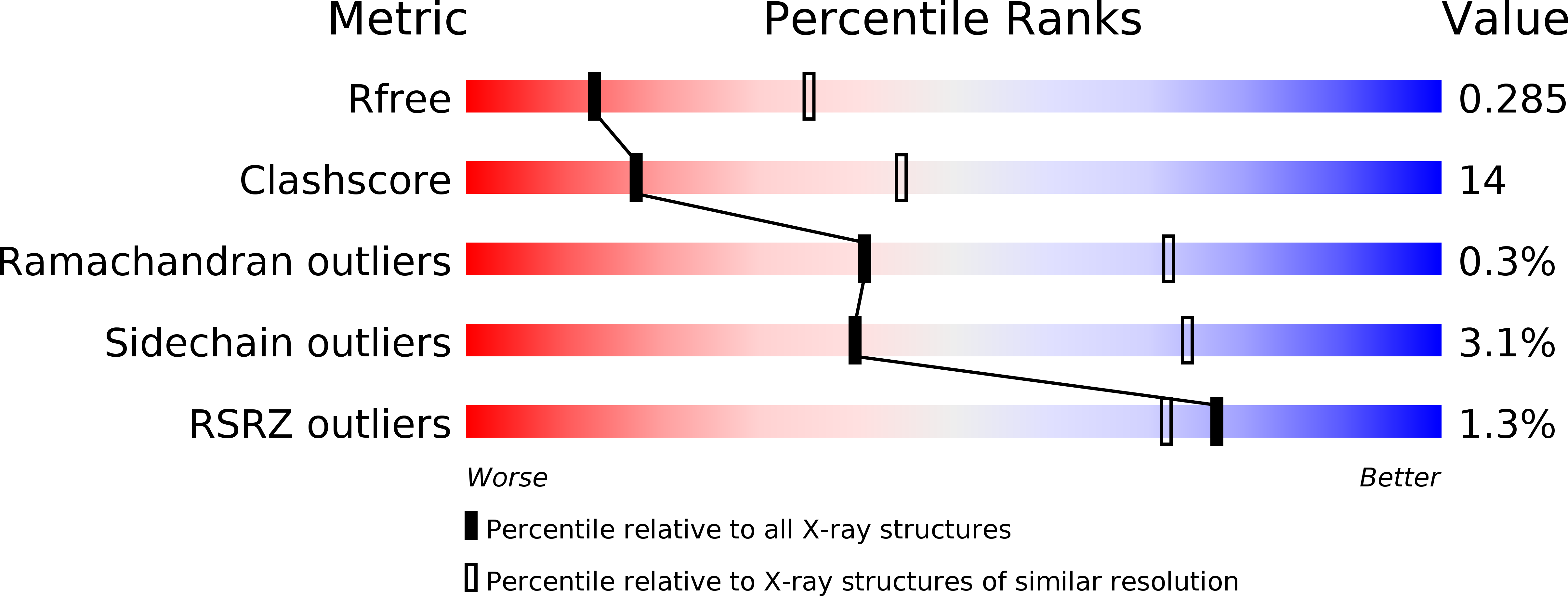
Resolution:
2.80 Å
R-Value Free:
0.27
R-Value Work:
0.23
R-Value Observed:
0.23
Space Group:
P 65
Macromolecular Entities
Polymer Type:polypeptide(L)
Molecule:Regulatory protein
Gene (Uniprot):esp1396IC
Chain IDs:A, B, C, D
Chain Length:82
Number of Molecules:4
Biological Source:Enterobacter sp. RFL1396
Polymer Type:polydeoxyribonucleotide
Molecule:35-MER DNA
Chain IDs:E
Chain Length:35
Number of Molecules:1
Biological Source:synthetic construct
Polymer Type:polydeoxyribonucleotide
Molecule:35-MER DNA
Chain IDs:F
Chain Length:35
Number of Molecules:1
Biological Source:synthetic construct
Ligand Molecules
Primary Citation
Radiation damage to nucleoprotein complexes in macromolecular crystallography.
J.Synchrotron Radiat. 22 213 224 (2015)
PMID: 25723923 DOI: 10.1107/S1600577514026289

Abstact

Significant progress has been made in macromolecular crystallography over recent years in both the understanding and mitigation of X-ray induced radiation damage when collecting diffraction data from crystalline proteins. In contrast, despite the large field that is productively engaged in the study of radiation chemistry of nucleic acids, particularly of DNA, there are currently very few X-ray crystallographic studies on radiation damage mechanisms in nucleic acids. Quantitative comparison of damage to protein and DNA crystals separately is challenging, but many of the issues are circumvented by studying pre-formed biological nucleoprotein complexes where direct comparison of each component can be made under the same controlled conditions. Here a model protein-DNA complex C.Esp1396I is employed to investigate specific damage mechanisms for protein and DNA in a biologically relevant complex over a large dose range (2.07-44.63 MGy). In order to allow a quantitative analysis of radiation damage sites from a complex series of macromolecular diffraction data, a computational method has been developed that is generally applicable to the field. Typical specific damage was observed for both the protein on particular amino acids and for the DNA on, for example, the cleavage of base-sugar N1-C and sugar-phosphate C-O bonds. Strikingly the DNA component was determined to be far more resistant to specific damage than the protein for the investigated dose range. At low doses the protein was observed to be susceptible to radiation damage while the DNA was far more resistant, damage only being observed at significantly higher doses.

Legend

Protein

Chemical

Disease

Primary Citation of related structures