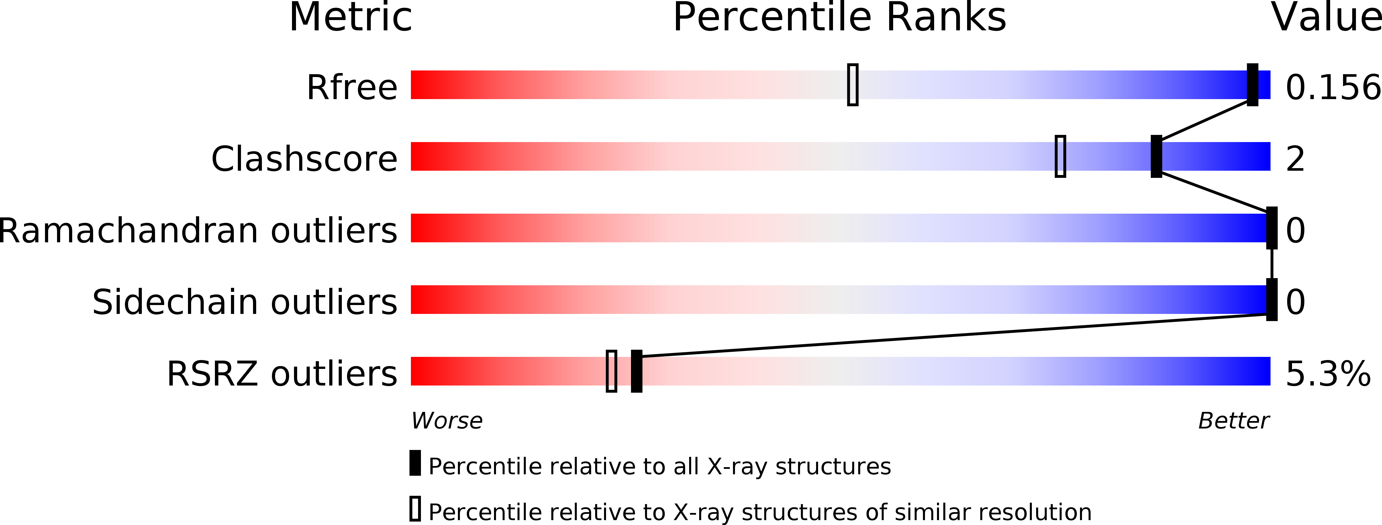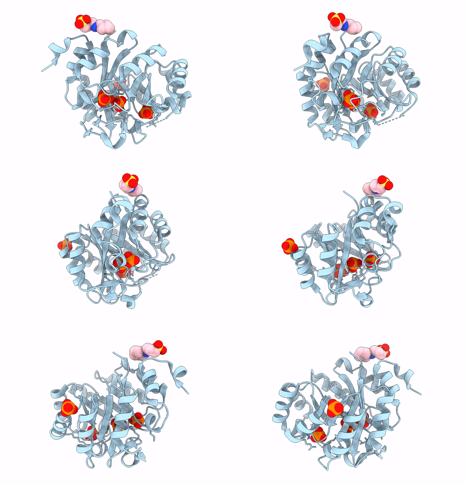
4X2R image
Deposition Date 2014-11-26
Release Date 2014-12-24
Last Version Date 2023-09-27
Entry Detail
PDB ID:
4X2R
Keywords:
Title:
Crystal structure of PriA from Actinomyces urogenitalis
Biological Source:
Source Organism:
Host Organism:
Method Details:
Experimental Method:
Resolution:
1.05 Å
R-Value Free:
0.14
R-Value Work:
0.12
R-Value Observed:
0.12
Space Group:
P 41
Macromolecular Entities
Structures with similar UniProt ID
Protein Blast
Polymer Type:polypeptide(L)
Molecule:1-(5-phosphoribosyl)-5-[(5-phosphoribosylamino)methylideneamino] imidazole-4-carboxamide isomerase
Gene (Uniprot):hisA
Chain IDs:A
Chain Length:250
Number of Molecules:1
Biological Source:Actinomyces urogenitalis DSM 15434
Primary Citation

Abstact

The connection between gene loss and the functional adaptation of retained proteins is still poorly understood. We apply phylogenomics and metabolic modeling to detect bacterial species that are evolving by gene loss, with the finding that Actinomycetaceae genomes from human cavities are undergoing sizable reductions, including loss of L-histidine and L-tryptophan biosynthesis. We observe that the dual-substrate phosphoribosyl isomerase A or priA gene, at which these pathways converge, appears to coevolve with the occurrence of trp and his genes. Characterization of a dozen PriA homologs shows that these enzymes adapt from bifunctionality in the largest genomes, to a monofunctional, yet not necessarily specialized, inefficient form in genomes undergoing reduction. These functional changes are accomplished via mutations, which result from relaxation of purifying selection, in residues structurally mapped after sequence and X-ray structural analyses. Our results show how gene loss can drive the evolution of substrate specificity from retained enzymes.

Legend

Protein

Chemical

Disease

Primary Citation of related structures