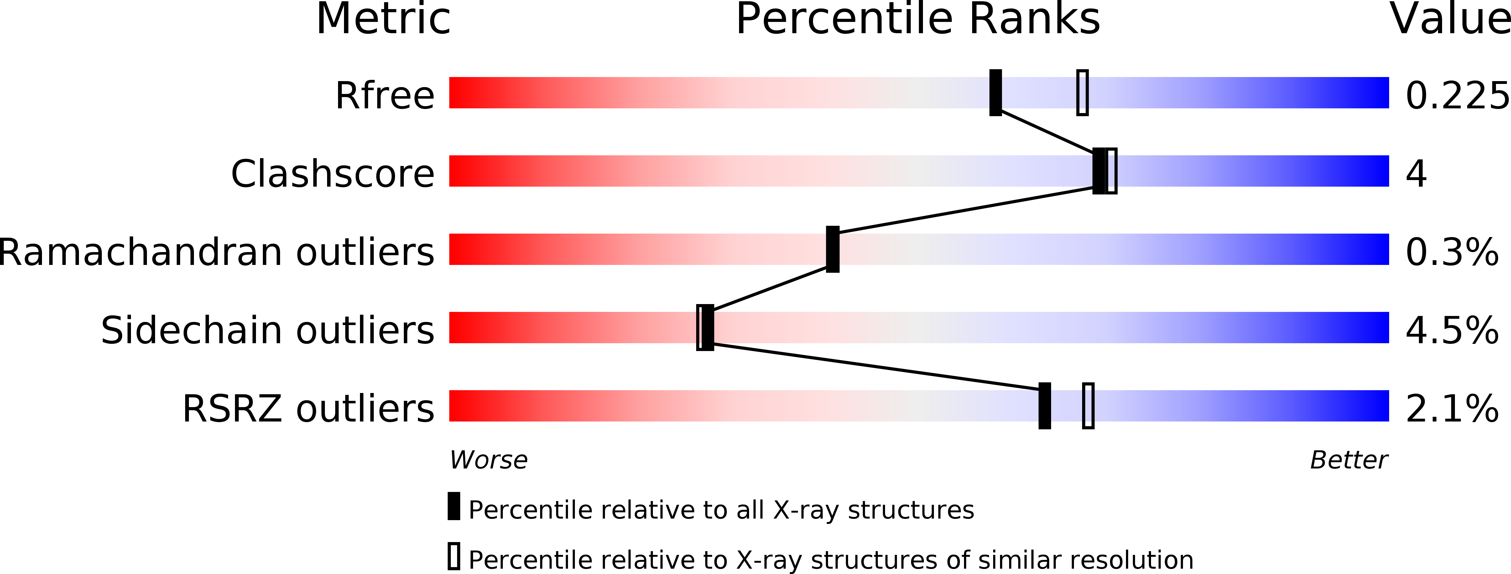
4WYS image
Deposition Date 2014-11-18
Release Date 2015-10-07
Last Version Date 2023-11-08
Entry Detail
PDB ID:
4WYS
Keywords:
Title:
Crystal structure of thiolase from Escherichia coli
Biological Source:
Source Organism:
Host Organism:
Method Details:
Experimental Method:
Resolution:
2.10 Å
R-Value Free:
0.21
R-Value Work:
0.16
R-Value Observed:
0.17
Space Group:
P 21 21 21
Macromolecular Entities
Structures with similar UniProt ID
Protein Blast
Polymer Type:polypeptide(L)
Molecule:Acetyl-CoA acetyltransferase
Gene (Uniprot):atoB
Chain IDs:A, B, C, D
Chain Length:405
Number of Molecules:4
Biological Source:Escherichia coli
Primary Citation
Redox-switch regulatory mechanism of thiolase from Clostridium acetobutylicum
Nat Commun 6 8410 8410 (2015)
PMID: 26391388 DOI: 10.1038/ncomms9410

Abstact

Thiolase is the first enzyme catalysing the condensation of two acetyl-coenzyme A (CoA) molecules to form acetoacetyl-CoA in a dedicated pathway towards the biosynthesis of n-butanol, an important solvent and biofuel. Here we elucidate the crystal structure of Clostridium acetobutylicum thiolase (CaTHL) in its reduced/oxidized states. CaTHL, unlike those from other aerobic bacteria such as Escherichia coli and Zoogloea ramegera, is regulated by the redox-switch modulation through reversible disulfide bond formation between two catalytic cysteine residues, Cys88 and Cys378. When CaTHL is overexpressed in wild-type C. acetobutylicum, butanol production is reduced due to the disturbance of acidogenic to solventogenic shift. The CaTHL(V77Q/N153Y/A286K) mutant, which is not able to form disulfide bonds, exhibits higher activity than wild-type CaTHL, and enhances butanol production upon overexpression. On the basis of these results, we suggest that CaTHL functions as a key enzyme in the regulation of the main metabolism of C. acetobutylicum through a redox-switch regulatory mechanism.

Legend

Protein

Chemical

Disease

Primary Citation of related structures