Deposition Date
2014-11-05
Release Date
2015-09-23
Last Version Date
2023-09-27
Entry Detail
PDB ID:
4WVG
Keywords:
Title:
Crystal structure of the Type-I signal peptidase from Staphylococcus aureus (SpsB).
Biological Source:
Source Organism(s):
Escherichia coli K-12 (Taxon ID: 83333)
Staphylococcus aureus subsp. aureus str. Newman (Taxon ID: 426430)
Staphylococcus aureus subsp. aureus str. Newman (Taxon ID: 426430)
Expression System(s):
Method Details:
Experimental Method:
Resolution:
2.05 Å
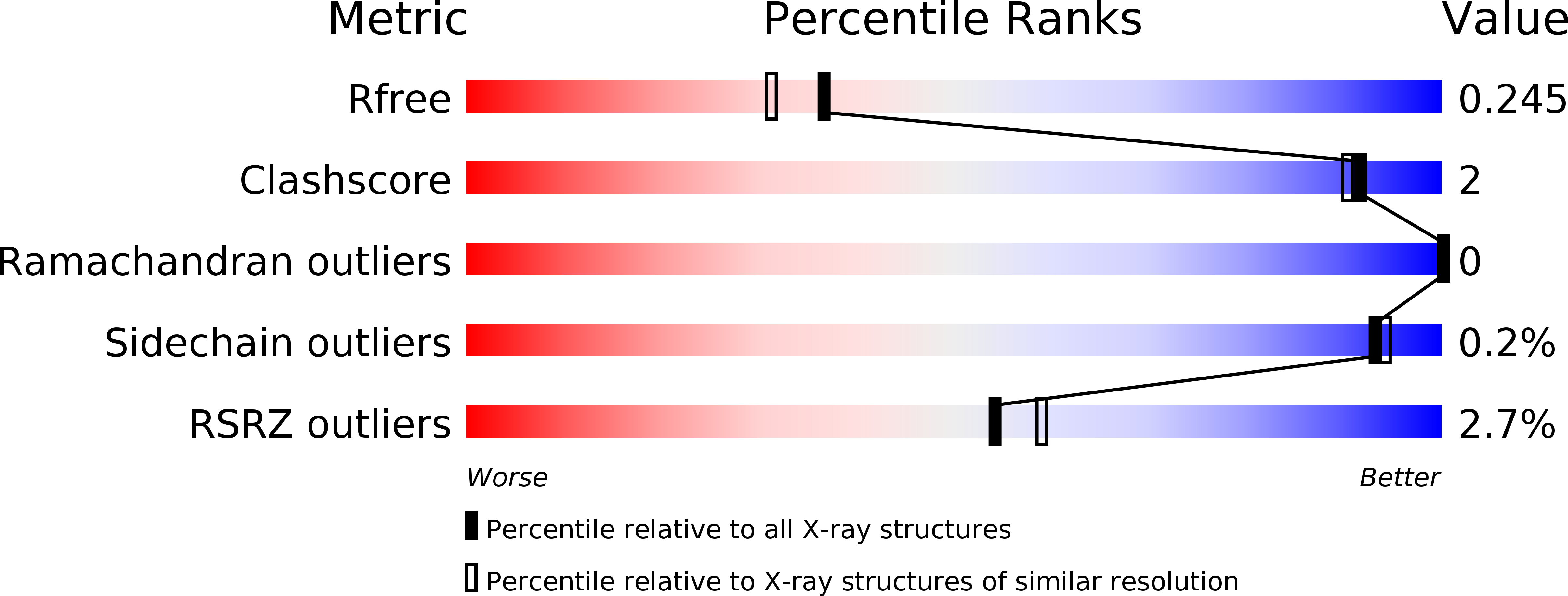
R-Value Free:
0.24
R-Value Work:
0.18
R-Value Observed:
0.19
Space Group:
P 1 21 1