4WUZ image
Deposition Date 2014-11-04
Release Date 2014-11-19
Last Version Date 2023-12-27
Entry Detail
PDB ID:
4WUZ
Keywords:
Title:
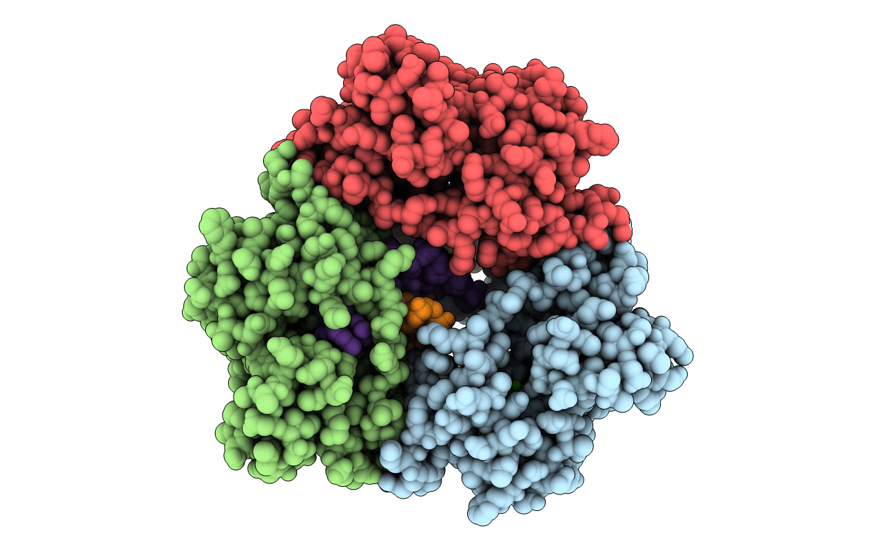
Crystal structure of lambda exonuclease in complex with DNA and Ca2+
Biological Source:
Source Organism:
Host Organism:
Method Details:
Experimental Method:
Resolution:
2.38 Å
R-Value Free:
0.31
R-Value Work:
0.23
R-Value Observed:
0.23
Space Group:
P 65
Macromolecular Entities
Polymer Type:polypeptide(L)
Molecule:Exonuclease
Gene (Uniprot):exo
Chain IDs:A, B, C
Chain Length:229
Number of Molecules:3
Biological Source:Enterobacteria phage lambda
Polymer Type:polydeoxyribonucleotide
Molecule:DNA (5'-D(*TP*T*TP*CP*GP*GP*TP*AP*CP*AP*GP*TP*AP*G)-3')
Chain IDs:D
Chain Length:14
Number of Molecules:1
Biological Source:synthetic
Polymer Type:polydeoxyribonucleotide
Molecule:DNA (5'-D(P*AP*GP*CP*TP*AP*CP*TP*GP*TP*AP*CP*CP*GP*A)-3')
Chain IDs:E
Chain Length:14
Number of Molecules:1
Biological Source:synthetic
Primary Citation
Crystal Structure of lambda Exonuclease in Complex with DNA and Ca(2+).
Biochemistry 53 7415 7425 (2014)
PMID: 25370446 DOI: 10.1021/bi501155q

Abstact

Bacteriophage λ exonuclease (λexo) is a ring-shaped homotrimer that resects double-stranded DNA ends in the 5'-3' direction to generate a long 3'-overhang that is a substrate for recombination. λexo is a member of the type II restriction endonuclease-like superfamily of proteins that use a Mg(2+)-dependent mechanism for nucleotide cleavage. A previous structure of λexo in complex with DNA and Mg(2+) was determined using a nuclease defective K131A variant to trap a stable complex. This structure revealed the detailed coordination of the two active site Mg(2+) ions but did not show the interactions involving the side chain of the conserved active site Lys-131 residue. Here, we have determined the crystal structure of wild-type (WT) λexo in complex with the same DNA substrate, but in the presence of Ca(2+) instead of Mg(2+). Surprisingly, there is only one Ca(2+) bound in the active site, near the position of Mg(A) in the structure with Mg(2+). The scissile phosphate is displaced by 2.2 Å relative to its position in the structure with Mg(2+), and the network of interactions involving the attacking water molecule is broken. Thus, the structure does not represent a catalytic configuration. However, the crystal structure does show clear electron density for the side chain of Lys-131, which is held in place by interactions with Gln-157 and Glu-129. By combining the K131A-Mg(2+) and WT-Ca(2+) structures, we constructed a composite model to show the likely interactions of Lys-131 during catalysis. The implications with regard to the catalytic mechanism are discussed.

Legend

Protein

Chemical

Disease

Primary Citation of related structures