Deposition Date
2014-10-31
Release Date
2015-04-08
Last Version Date
2024-01-10
Entry Detail
PDB ID:
4WUD
Keywords:
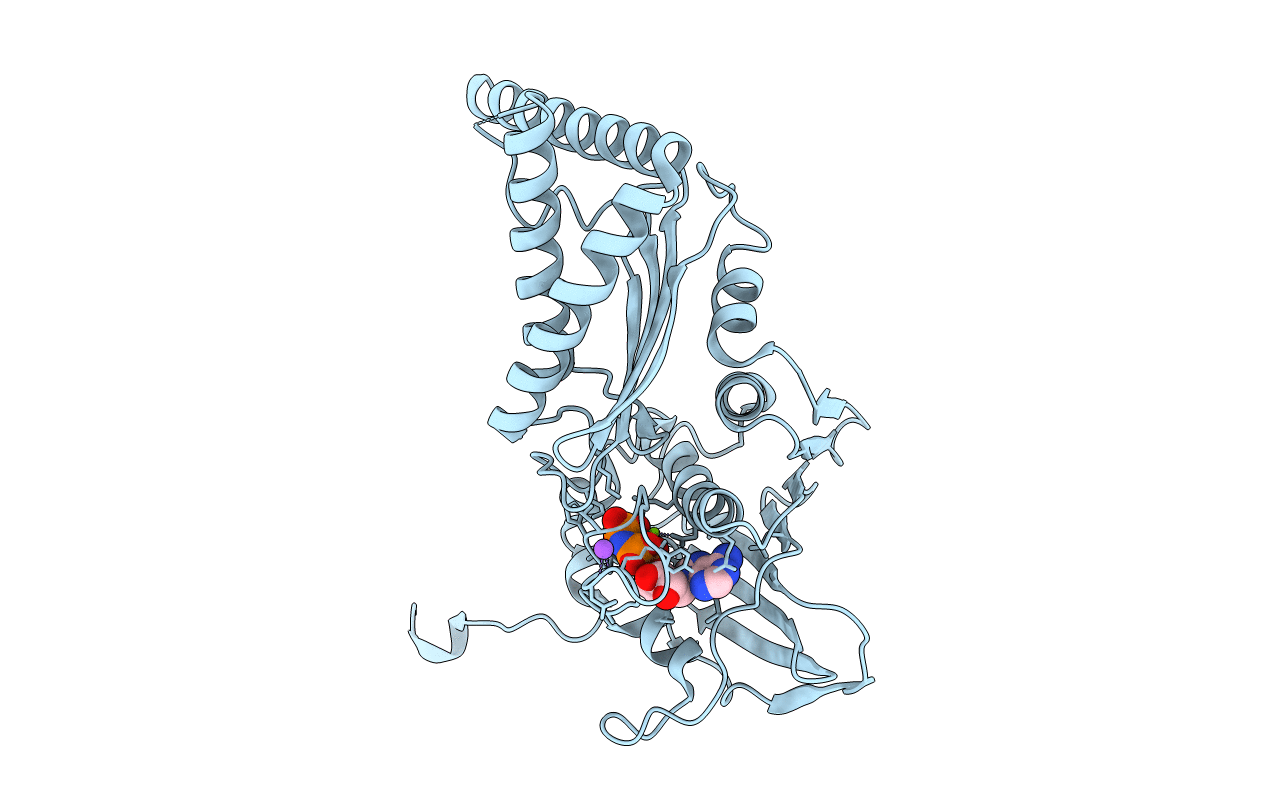
Title:
N-terminal 43 kDa fragment of the E. coli DNA gyrase B subunit grown from no salt condition
Biological Source:
Source Organism(s):
Escherichia coli strain K-12 (Taxon ID: 83333)
Expression System(s):
Method Details:
Experimental Method:
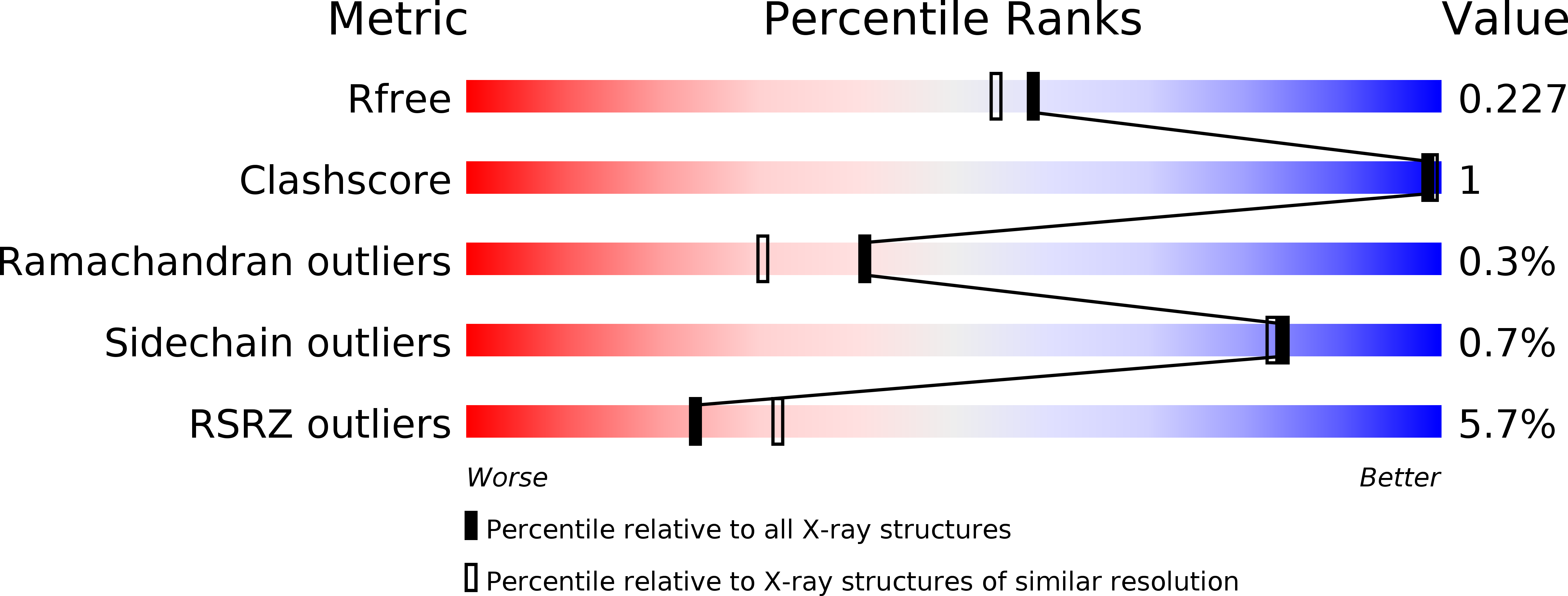
Resolution:
1.95 Å
R-Value Free:
0.21
R-Value Work:
0.18
R-Value Observed:
0.18
Space Group:
C 2 2 21