4WU0 image
Deposition Date 2014-10-30
Release Date 2015-07-08
Last Version Date 2023-09-27
Entry Detail
PDB ID:
4WU0
Keywords:
Title:
Structural Analysis of C. acetobutylicum ATCC 824 Glycoside Hydrolase From Family 105
Biological Source:
Source Organism:
Host Organism:
Method Details:
Experimental Method:
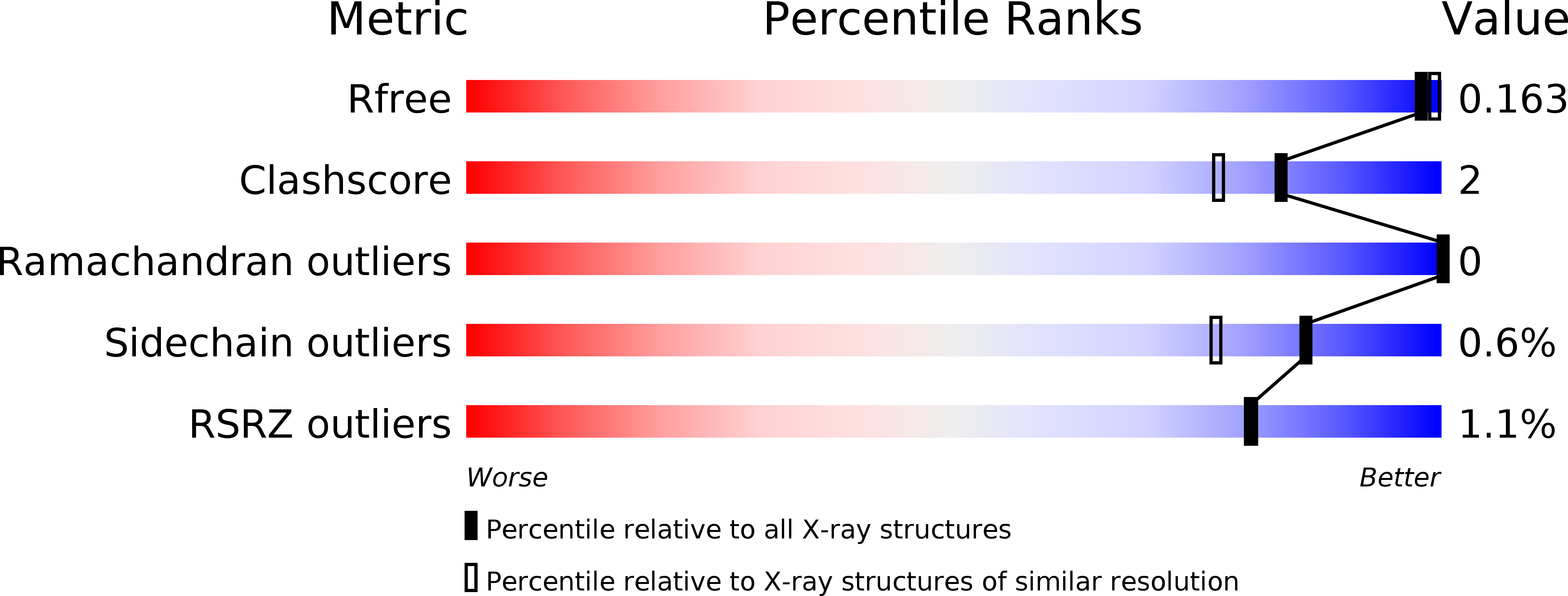
Resolution:
1.60 Å
R-Value Free:
0.16
R-Value Work:
0.13
R-Value Observed:
0.13
Space Group:
P 21 21 21
Macromolecular Entities
Structures with similar UniProt ID
Protein Blast
Polymer Type:polypeptide(L)
Molecule:Similar to yteR (Bacilus subtilis)
Gene (Uniprot):CA_C0359
Chain IDs:A, B
Chain Length:361
Number of Molecules:2
Biological Source:Clostridium acetobutylicum
Primary Citation
Structural analysis of Clostridium acetobutylicum ATCC 824 glycoside hydrolase from CAZy family GH105.
Acta Crystallogr.,Sect.F 71 1100 1108 (2015)
PMID: 26249707 DOI: 10.1107/S2053230X15012121

Abstact

Clostridium acetobutylicum ATCC 824 gene CA_C0359 encodes a putative unsaturated rhamnogalacturonyl hydrolase (URH) with distant amino-acid sequence homology to YteR of Bacillus subtilis strain 168. YteR, like other URHs, has core structural homology to unsaturated glucuronyl hydrolases, but hydrolyzes the unsaturated disaccharide derivative of rhamnogalacturonan I. The crystal structure of the recombinant CA_C0359 protein was solved to 1.6 Å resolution by molecular replacement using the phase information of the previously reported structure of YteR (PDB entry 1nc5) from Bacillus subtilis strain 168. The YteR-like protein is a six-α-hairpin barrel with two β-sheet strands and a small helix overlaying the end of the hairpins next to the active site. The protein has low primary protein sequence identity to YteR but is structurally similar. The two tertiary structures align with a root-mean-square deviation of 1.4 Å and contain a highly conserved active pocket. There is a conserved aspartic acid residue in both structures, which has been shown to be important for hydration of the C=C bond during the release of unsaturated galacturonic acid by YteR. A surface electrostatic potential comparison of CA_C0359 and proteins from CAZy families GH88 and GH105 reveals the make-up of the active site to be a combination of the unsaturated rhamnogalacturonyl hydrolase and the unsaturated glucuronyl hydrolase from Bacillus subtilis strain 168. Structural and electrostatic comparisons suggests that the protein may have a slightly different substrate specificity from that of YteR.

Legend

Protein

Chemical

Disease

Primary Citation of related structures
Feedback Form
Name
Email
Institute
Feedback