Deposition Date
2014-10-30
Release Date
2015-08-12
Last Version Date
2024-10-23
Entry Detail
PDB ID:
4WTS
Keywords:
Title:
Active-site mutant of Rhizomucor miehei beta-1,3-glucanosyltransferase in complex with laminaritriose
Biological Source:
Source Organism:
Rhizomucor miehei CAU432 (Taxon ID: 1031333)
Host Organism:
Method Details:
Experimental Method:
Resolution:
2.30 Å
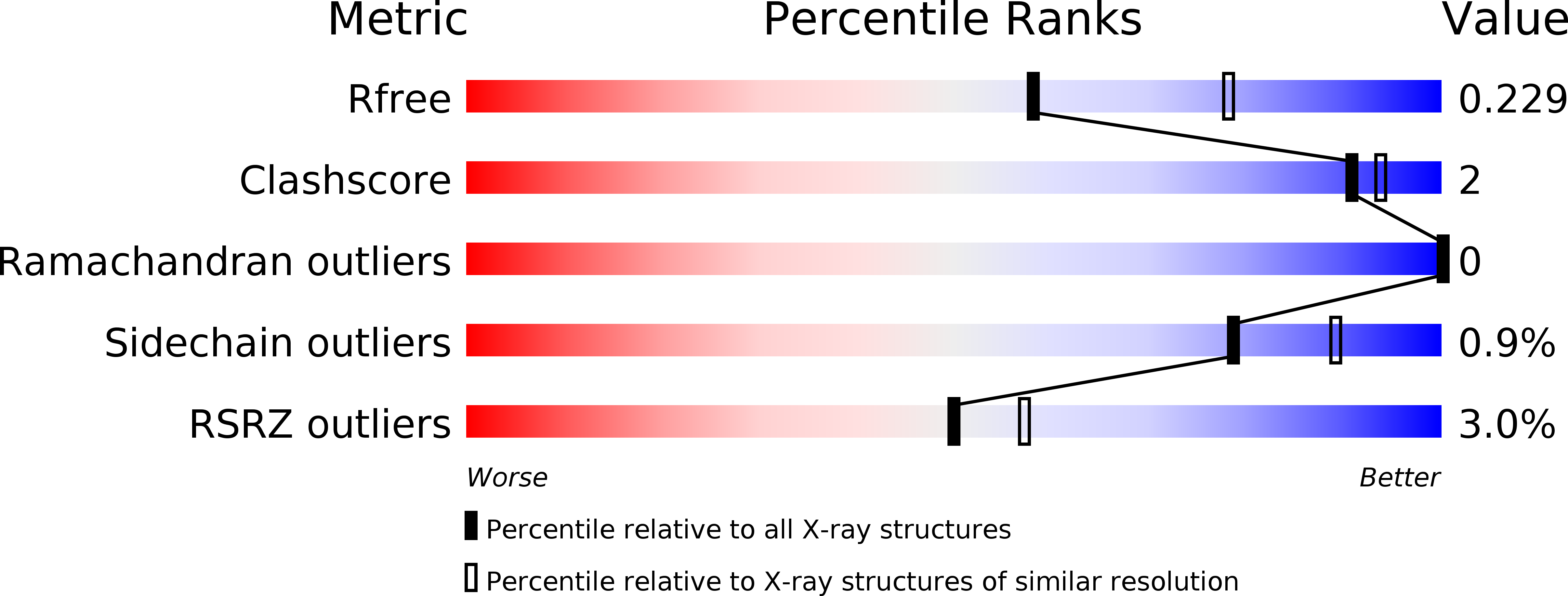
R-Value Free:
0.24
R-Value Work:
0.21
R-Value Observed:
0.21
Space Group:
P 21 21 21