
Deposition Date
2014-10-25
Release Date
2015-07-15
Last Version Date
2023-09-27
Entry Detail
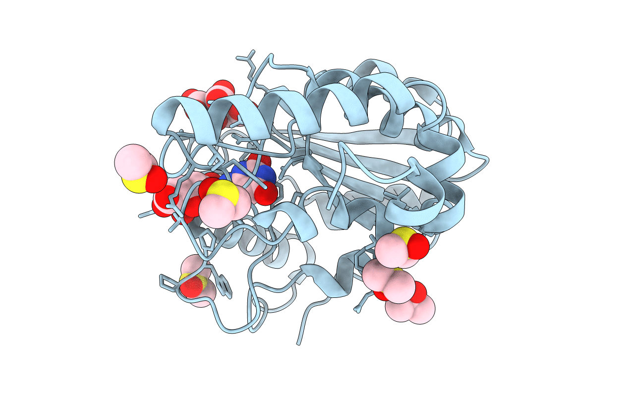
PDB ID:
4WS2
Keywords:
Title:
Crystal structure of Mycobacterium tuberculosis uracil-DNA glycosylase in complex with 6-aminouracil, Form I
Biological Source:
Source Organism(s):
Mycobacterium tuberculosis (Taxon ID: 83332)
Expression System(s):
Method Details:
Experimental Method:
Resolution:
1.13 Å
R-Value Free:
0.15
R-Value Work:
0.12
R-Value Observed:
0.12
Space Group:
P 1 21 1
