4WRI image
Deposition Date 2014-10-24
Release Date 2015-05-27
Last Version Date 2025-04-09
Entry Detail
PDB ID:
4WRI
Title:
Crystal structure of okadaic acid binding protein 2.1
Biological Source:
Source Organism:
Host Organism:
Method Details:
Experimental Method:
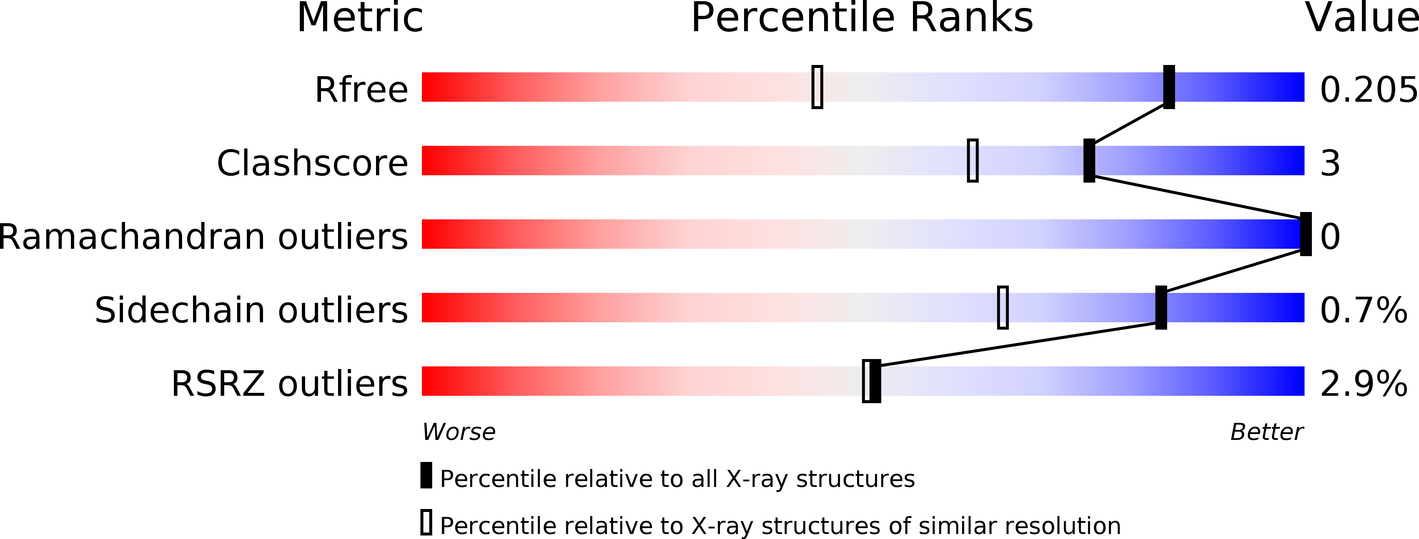
Resolution:
1.40 Å
R-Value Free:
0.20
R-Value Work:
0.17
R-Value Observed:
0.17
Space Group:
C 2 2 21
Macromolecular Entities
Structures with similar UniProt ID
Protein Blast
Polymer Type:polypeptide(L)
Molecule:Okadaic acid binding protein 2-alpha
Gene (Uniprot):OABP2A
Chain IDs:A
Chain Length:192
Number of Molecules:1
Biological Source:Halichondria okadai
Modified Residue
Compound ID Chain ID Parent Comp ID Details 2D Image
MLY A LYS modified residue
Ligand Molecules
Primary Citation
Crystal Structure of Okadaic Acid Binding Protein 2.1: A Sponge Protein Implicated in Cytotoxin Accumulation
Chembiochem 16 1435 1439 (2015)
PMID: 25965326 DOI: 10.1002/cbic.201500141

Abstact

Okadaic acid (OA) is a marine polyether cytotoxin that was first isolated from the marine sponge Halichondria okadai. OA is a potent inhibitor of protein serine/threonine phosphatases (PP) 1 and 2A, and the structural basis of phosphatase inhibition has been well investigated. However, the role and mechanism of OA retention in the marine sponge have remained elusive. We have solved the crystal structure of okadaic acid binding protein 2.1 (OABP2.1) isolated from H. okadai; it has strong affinity for OA and limited sequence homology to other proteins. The structure revealed that OABP2.1 consists of two α-helical domains, with the OA molecule deeply buried inside the protein. In addition, the global fold of OABP2.1 was unexpectedly similar to that of aequorin, a jellyfish photoprotein. The presence of structural homologues suggested that, by using similar protein scaffolds, marine invertebrates have developed diverse survival systems adapted to their living environments.

Legend

Protein

Chemical

Disease

Primary Citation of related structures