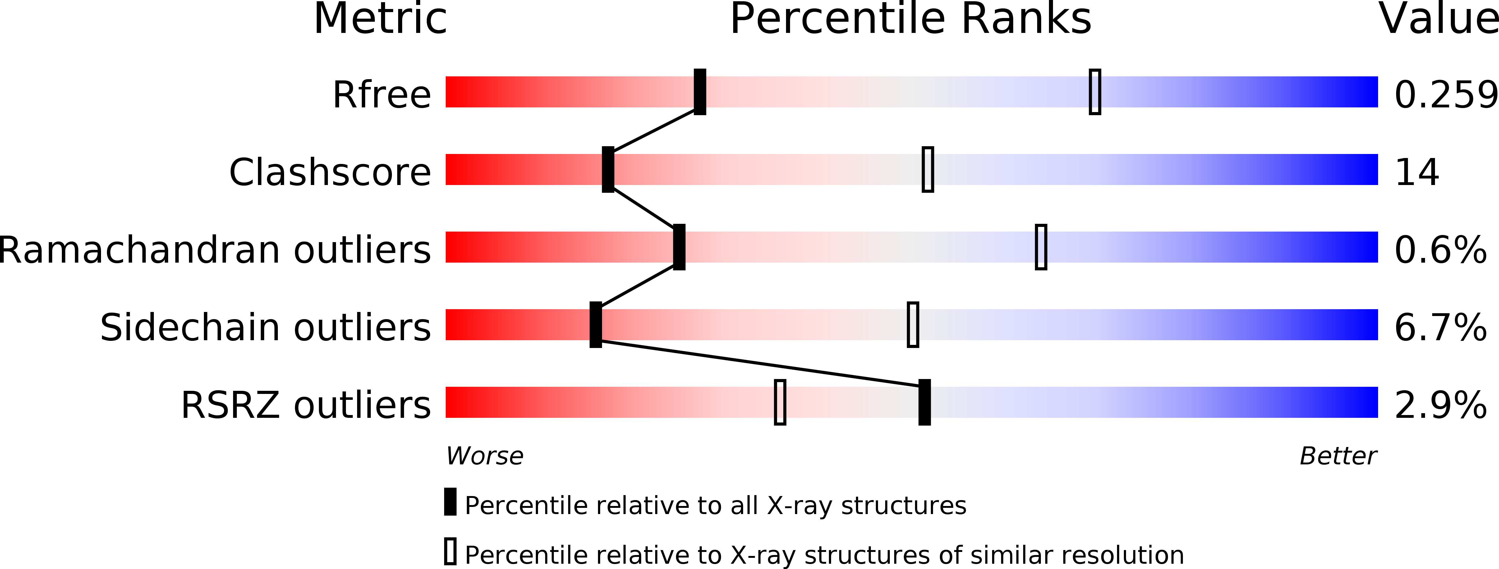
4WQO image
Deposition Date 2014-10-22
Release Date 2015-03-04
Last Version Date 2023-09-27
Entry Detail
PDB ID:
4WQO
Keywords:
Title:
Structure of VHL-EloB-EloC-Cul2
Biological Source:
Source Organism:
Homo sapiens (Taxon ID: 9606)
Host Organism:
Method Details:
Experimental Method:
Resolution:
3.20 Å
R-Value Free:
0.24
R-Value Work:
0.22
R-Value Observed:
0.22
Space Group:
P 32 2 1
Macromolecular Entities
Structures with similar UniProt ID
Protein Blast
Polymer Type:polypeptide(L)
Molecule:Von Hippel-Lindau disease tumor suppressor
Gene (Uniprot):VHL
Chain IDs:A
Chain Length:233
Number of Molecules:1
Biological Source:Homo sapiens
Structures with similar UniProt ID
Protein Blast
Polymer Type:polypeptide(L)
Molecule:Transcription elongation factor B polypeptide 2
Gene (Uniprot):ELOB
Chain IDs:B
Chain Length:118
Number of Molecules:1
Biological Source:Homo sapiens
Structures with similar UniProt ID
Protein Blast
Polymer Type:polypeptide(L)
Molecule:Transcription elongation factor B polypeptide 1
Gene (Uniprot):ELOC
Chain IDs:C
Chain Length:96
Number of Molecules:1
Biological Source:Homo sapiens
Structures with similar UniProt ID
Protein Blast
Polymer Type:polypeptide(L)
Molecule:Cullin-2
Gene (Uniprot):CUL2
Chain IDs:D
Chain Length:186
Number of Molecules:1
Biological Source:Homo sapiens
Ligand Molecules
Primary Citation
Insights into Cullin-RING E3 Ubiquitin Ligase Recruitment: Structure of the VHL-EloBC-Cul2 Complex.
Structure 23 441 449 (2015)
PMID: 25661653 DOI: 10.1016/j.str.2014.12.014

Abstact

The von Hippel-Lindau tumor suppressor protein (VHL) recruits a Cullin 2 (Cul2) E3 ubiquitin ligase to downregulate HIF-1α, an essential transcription factor for the hypoxia response. Mutations in VHL lead to VHL disease and renal cell carcinomas. Inhibition of this pathway to upregulate erythropoietin production is a promising new therapy to treat ischemia and chronic anemia. Here, we report the crystal structure of VHL bound to a Cul2 N-terminal domain, Elongin B, and Elongin C (EloC). Cul2 interacts with both the VHL BC box and cullin box and a novel EloC site. Comparison with other cullin E3 ligase structures shows that there is a conserved, yet flexible, cullin recognition module and that cullin selectivity is influenced by distinct electrostatic interactions. Our structure provides a structural basis for the study of the pathogenesis of VHL disease and rationale for the design of novel compounds that may modulate cullin-substrate receptor interactions.

Legend

Protein

Chemical

Disease

Primary Citation of related structures