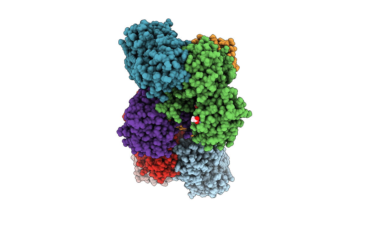
4WNC image
Deposition Date 2014-10-11
Release Date 2014-12-03
Last Version Date 2023-09-27
Entry Detail
PDB ID:
4WNC
Keywords:
Title:
Crystal structure of human wild-type GAPDH at 1.99 angstroms resolution
Biological Source:
Source Organism:
Homo sapiens (Taxon ID: 9606)
Host Organism:
Method Details:
Experimental Method:
Resolution:
1.99 Å
R-Value Free:
0.17
R-Value Work:
0.13
R-Value Observed:
0.13
Space Group:
P 1 21 1
Macromolecular Entities
Polymer Type:polypeptide(L)
Molecule:Glyceraldehyde-3-phosphate dehydrogenase
Gene (Uniprot):GAPDH
Chain IDs:A (auth: O), B (auth: A), C (auth: B), D (auth: C), E (auth: D), F (auth: E), G (auth: F), H (auth: G)
Chain Length:335
Number of Molecules:8
Biological Source:Homo sapiens
Primary Citation
A Dimer Interface Mutation in Glyceraldehyde-3-Phosphate Dehydrogenase Regulates Its Binding to AU-rich RNA.
J.Biol.Chem. 290 1770 1785 (2015)
PMID: 25451934 DOI: 10.1074/jbc.M114.618165

Abstact

Glyceraldehyde-3-phosphate dehydrogenase (GAPDH) is an enzyme best known for its role in glycolysis. However, extra-glycolytic functions of GAPDH have been described, including regulation of protein expression via RNA binding. GAPDH binds to numerous adenine-uridine rich elements (AREs) from various mRNA 3'-untranslated regions in vitro and in vivo despite its lack of a canonical RNA binding motif. How GAPDH binds to these AREs is still unknown. Here we discovered that GAPDH binds with high affinity to the core ARE from tumor necrosis factor-α mRNA via a two-step binding mechanism. We demonstrate that a mutation at the GAPDH dimer interface impairs formation of the second RNA-GAPDH complex and leads to changes in the RNA structure. We investigated the effect of this interfacial mutation on GAPDH oligomerization by crystallography, small-angle x-ray scattering, nano-electrospray ionization native mass spectrometry, and hydrogen-deuterium exchange mass spectrometry. We show that the mutation does not significantly affect GAPDH tetramerization as previously proposed. Instead, the mutation promotes short-range and long-range dynamic changes in regions located at the dimer and tetramer interface and in the NAD(+) binding site. These dynamic changes are localized along the P axis of the GAPDH tetramer, suggesting that this region is important for RNA binding. Based on our results, we propose a model for sequential GAPDH binding to RNA via residues located at the dimer and tetramer interfaces.

Legend

Protein

Chemical

Disease

Primary Citation of related structures