4WMY image
Deposition Date 2014-10-09
Release Date 2015-07-01
Last Version Date 2024-11-13
Entry Detail
PDB ID:
4WMY
Title:
Structure of Human intelectin-1 in complex with allyl-beta-galactofuranose
Biological Source:
Source Organism:
Homo sapiens (Taxon ID: 9606)
Host Organism:
Method Details:
Experimental Method:
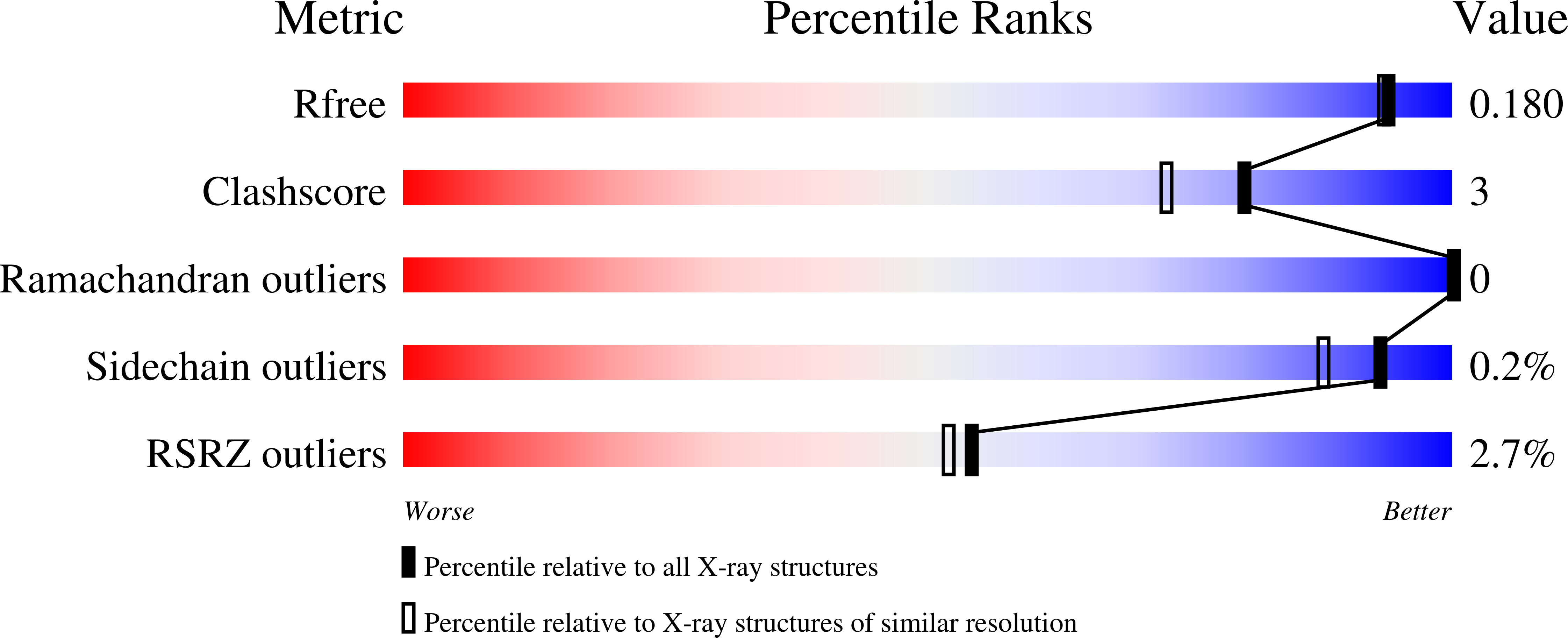
Resolution:
1.60 Å
R-Value Free:
0.17
R-Value Work:
0.15
R-Value Observed:
0.15
Space Group:
P 21 3
Macromolecular Entities
Structures with similar UniProt ID
Protein Blast
Polymer Type:polypeptide(L)
Molecule:Intelectin-1
Gene (Uniprot):ITLN1
Chain IDs:A, B
Chain Length:306
Number of Molecules:2
Biological Source:Homo sapiens
Primary Citation
Recognition of microbial glycans by human intelectin-1.
Nat.Struct.Mol.Biol. 22 603 610 (2015)
PMID: 26148048 DOI: 10.1038/nsmb.3053

Abstact

The glycans displayed on mammalian cells can differ markedly from those on microbes. Such differences could, in principle, be 'read' by carbohydrate-binding proteins, or lectins. We used glycan microarrays to show that human intelectin-1 (hIntL-1) does not bind known human glycan epitopes but does interact with multiple glycan epitopes found exclusively on microbes: β-linked D-galactofuranose (β-Galf), D-phosphoglycerol-modified glycans, heptoses, D-glycero-D-talo-oct-2-ulosonic acid (KO) and 3-deoxy-D-manno-oct-2-ulosonic acid (KDO). The 1.6-Å-resolution crystal structure of hIntL-1 complexed with β-Galf revealed that hIntL-1 uses a bound calcium ion to coordinate terminal exocyclic 1,2-diols. N-acetylneuraminic acid (Neu5Ac), a sialic acid widespread in human glycans, has an exocyclic 1,2-diol but does not bind hIntL-1, probably owing to unfavorable steric and electronic effects. hIntL-1 marks only Streptococcus pneumoniae serotypes that display surface glycans with terminal 1,2-diol groups. This ligand selectivity suggests that hIntL-1 functions in microbial surveillance.

Legend

Protein

Chemical

Disease

Primary Citation of related structures