4WL2 image
Deposition Date 2014-10-06
Release Date 2015-10-07
Last Version Date 2024-10-16
Entry Detail
PDB ID:
4WL2
Keywords:
Title:
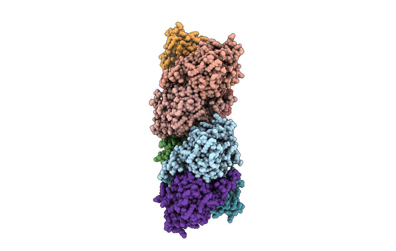
Structure of penicillin V acylase from Pectobacterium atrosepticum
Biological Source:
Source Organism:
Host Organism:
Method Details:
Experimental Method:
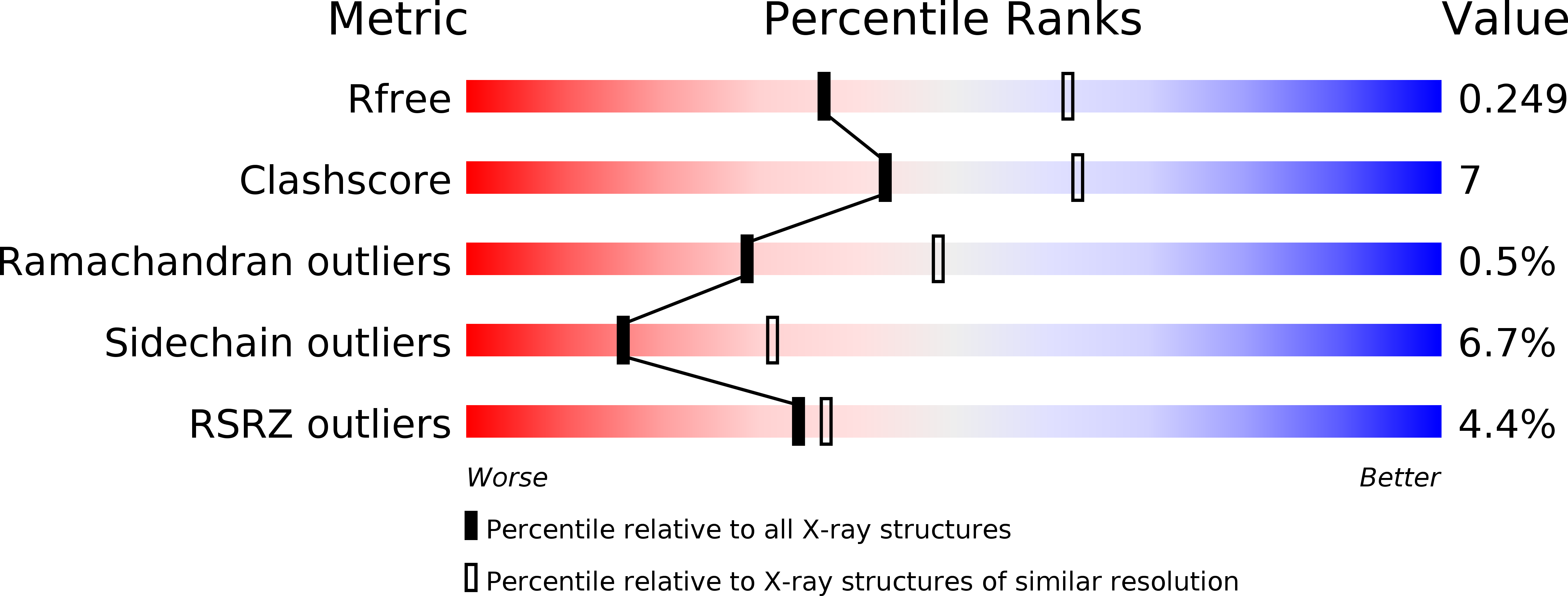
Resolution:
2.50 Å
R-Value Free:
0.24
R-Value Work:
0.21
R-Value Observed:
0.21
Space Group:
P 21 21 21
Macromolecular Entities
Polymer Type:polypeptide(L)
Molecule:Putative exported choloylglycine hydrolase
Gene (Uniprot):pva
Chain IDs:A, B, C, D, E, F, G, H
Chain Length:355
Number of Molecules:8
Biological Source:Pectobacterium atrosepticum SCRI1043
Primary Citation
Structural analysis of a penicillin V acylase from Pectobacterium atrosepticum confirms the importance of two Trp residues for activity and specificity
J.Struct.Biol. 193 85 94 (2016)
PMID: 26707624 DOI: 10.1016/j.jsb.2015.12.008

Abstact

Penicillin V acylases (PVA) catalyze the deacylation of the beta-lactam antibiotic phenoxymethylpenicillin (Pen V). They are members of the Ntn hydrolase family and possess an N-terminal cysteine as the main catalytic nucleophile residue. They form the evolutionarily related cholylglycine hydrolase (CGH) group which includes bile salt hydrolases (BSH) responsible for bile deconjugation. Even though a few PVA and BSH structures have been reported, no structure of a functional PVA from Gram-negative bacteria is available. Here, we report the crystal structure of a highly active PVA from Gram-negative Pectobacterium atrosepticum (PaPVA) at 2.5Å resolution. Structural comparison with PVAs from Gram-positive bacteria revealed that PaPVA had a distinctive tetrameric structure and active site organization. In addition, mutagenesis of key active site residues and biochemical characterization of the resultant variants elucidated the role of these residues in substrate binding and catalysis. The importance of residue Trp23 and Trp87 side chains in binding and correct positioning of Pen V by PVAs was confirmed using mutagenesis and substrate docking with a 15ns molecular dynamics simulation. These results establish the unique nature of Gram-negative CGHs and necessitate further research about their substrate spectrum.

Legend

Protein

Chemical

Disease

Primary Citation of related structures