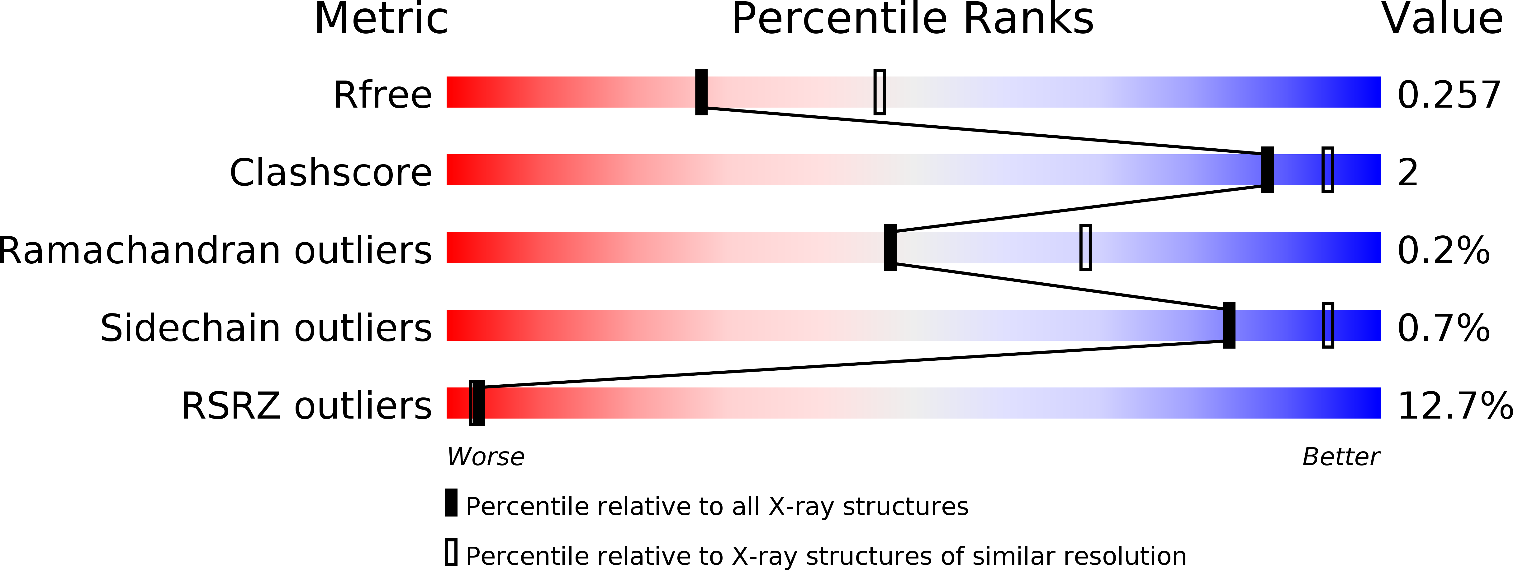
4WK2 image
Deposition Date 2014-10-01
Release Date 2014-12-03
Last Version Date 2024-11-20
Entry Detail
PDB ID:
4WK2
Title:
Metal Ion and Ligand Binding of Integrin
Biological Source:
Source Organism:
Homo sapiens (Taxon ID: 9606)
Host Organism:
Method Details:
Experimental Method:
Resolution:
2.50 Å
R-Value Free:
0.25
R-Value Work:
0.21
R-Value Observed:
0.21
Space Group:
P 21 21 21
Macromolecular Entities
Polymer Type:polypeptide(L)
Molecule:Integrin alpha-5
Gene (Uniprot):ITGA5
Mutations:I451V
Chain IDs:A
Chain Length:452
Number of Molecules:1
Biological Source:Homo sapiens
Polymer Type:polypeptide(L)
Molecule:Integrin beta-1
Gene (Uniprot):ITGB1
Chain IDs:B
Chain Length:445
Number of Molecules:1
Biological Source:Homo sapiens
Polymer Type:polypeptide(L)
Molecule:GLY-ARG-GLY-ASP-SER-PRO
Chain IDs:C
Chain Length:6
Number of Molecules:1
Biological Source:Homo sapiens
Primary Citation
Metal ion and ligand binding of integrin alpha 5 beta 1.
Proc.Natl.Acad.Sci.USA 111 17863 17868 (2014)
PMID: 25475857 DOI: 10.1073/pnas.1420645111

Abstact

Integrin α5β1 binds to an Arg-Gly-Asp (RGD) motif in its ligand fibronectin. We report high-resolution crystal structures of a four-domain α5β1 headpiece fragment, alone or with RGD peptides soaked into crystals, and RGD peptide affinity measurements. The headpiece crystallizes in a closed conformation essentially identical to that seen previously for α5β1 complexed with a Fab that allosterically inhibits ligand binding by stabilizing the closed conformation. Soaking experiments show that binding of cyclic RGD peptide with 20-fold higher affinity than a linear RGD peptide induces conformational change in the β1-subunit βI domain to a state that is intermediate between closed (low affinity) and open (high affinity). In contrast, binding of a linear RGD peptide induces no shape shifting. However, linear peptide binding induces shape shifting when Ca(2+) is depleted during soaking. Ca(2+) bound to the adjacent to metal ion-dependent adhesion site (ADMIDAS), at the locus of shape shifting, moves and decreases in occupancy, correlating with an increase in affinity for RGD measured when Ca(2+) is depleted. The results directly demonstrate that Ca(2+) binding to the ADMIDAS stabilizes integrins in the low-affinity, closed conformation. Comparisons in affinity between four-domain and six-domain headpiece constructs suggest that flexible integrin leg domains contribute to conformational equilibria. High-resolution views of the hybrid domain interface with the plexin-semaphorin-integrin (PSI) domain in different orientations show a ball-and-socket joint with a hybrid domain Arg side chain that rocks in a PSI domain socket lined with carbonyl oxygens.

Legend

Protein

Chemical

Disease

Primary Citation of related structures