4WIT image
Deposition Date 2014-09-26
Release Date 2014-11-12
Last Version Date 2024-05-08
Entry Detail
PDB ID:
4WIT
Keywords:
Title:
TMEM16 lipid scramblase in crystal form 2
Biological Source:
Source Organism:
Method Details:
Experimental Method:
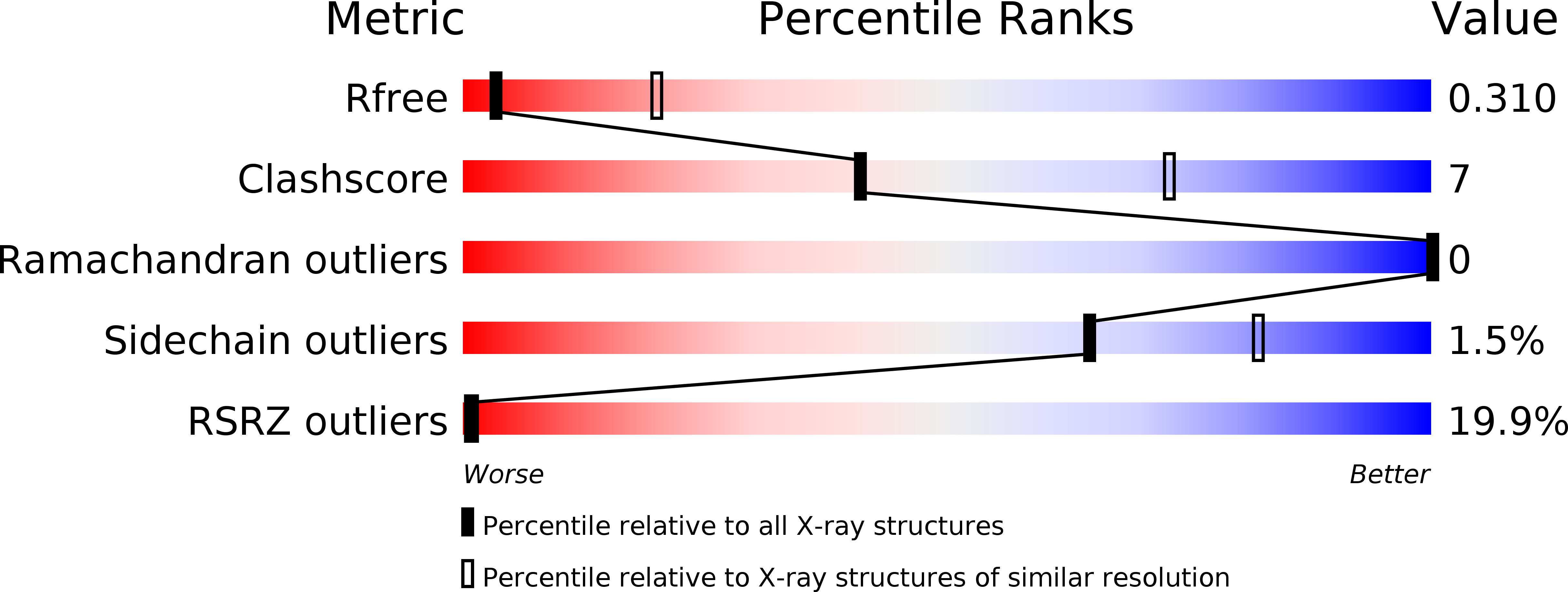
Resolution:
3.40 Å
R-Value Free:
0.29
R-Value Work:
0.24
R-Value Observed:
0.24
Space Group:
P 21 21 21
Macromolecular Entities
Structures with similar UniProt ID
Protein Blast
Polymer Type:polypeptide(L)
Molecule:Predicted protein
Gene (Uniprot):NECHADRAFT_66456
Chain IDs:A, B
Chain Length:735
Number of Molecules:2
Biological Source:Nectria haematococca
Ligand Molecules
Primary Citation
X-ray structure of a calcium-activated TMEM16 lipid scramblase.
Nature 516 207 212 (2014)
PMID: 25383531 DOI: 10.1038/nature13984

Abstact

The TMEM16 family of proteins, also known as anoctamins, features a remarkable functional diversity. This family contains the long sought-after Ca(2+)-activated chloride channels as well as lipid scramblases and cation channels. Here we present the crystal structure of a TMEM16 family member from the fungus Nectria haematococca that operates as a Ca(2+)-activated lipid scramblase. Each subunit of the homodimeric protein contains ten transmembrane helices and a hydrophilic membrane-traversing cavity that is exposed to the lipid bilayer as a potential site of catalysis. This cavity harbours a conserved Ca(2+)-binding site located within the hydrophobic core of the membrane. Mutations of residues involved in Ca(2+) coordination affect both lipid scrambling in N. haematococca TMEM16 and ion conduction in the Cl(-) channel TMEM16A. The structure reveals the general architecture of the family and its mode of Ca(2+) activation. It also provides insight into potential scrambling mechanisms and serves as a framework to unravel the conduction of ions in certain TMEM16 proteins.

Legend

Protein

Chemical

Disease

Primary Citation of related structures