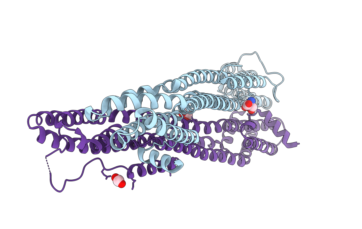
4WID image
Entry Detail
PDB ID:
4WID
Keywords:
Title:
Crystal structure of the immediate-early 1 protein (IE1) at 2.31 angstrom (tetragonal form after crystal dehydration)
Biological Source:
Source Organism:
Host Organism:
PDB Version:
Deposition Date:
2014-09-25
Release Date:
2014-10-29
Method Details:
Experimental Method:
Resolution:
2.31 Å
R-Value Free:
0.24
R-Value Work:
0.19
R-Value Observed:
0.20
Space Group:
P 43
Macromolecular Entities
Polymer Type:polypeptide(L)
Description:RhUL123
Chain IDs:A
Chain Length:365
Number of Molecules:1
Biological Source:Macacine herpesvirus 3
Polymer Type:polypeptide(L)
Description:RhUL123
Chain IDs:B
Chain Length:365
Number of Molecules:1
Biological Source:Macacine herpesvirus 3
Modified Residue
Compound ID Chain ID Parent Comp ID Details 2D Image
MLY A LYS modified residue
MLZ A LYS modified residue
Primary Citation
Crystal Structure of Cytomegalovirus IE1 Protein Reveals Targeting of TRIM Family Member PML via Coiled-Coil Interactions.
Plos Pathog. 10 e1004512 e1004512 (2014)
PMID: 25412268 DOI: 10.1371/journal.ppat.1004512

Abstact

PML nuclear bodies (PML-NBs) are enigmatic structures of the cell nucleus that act as key mediators of intrinsic immunity against viral pathogens. PML itself is a member of the E3-ligase TRIM family of proteins that regulates a variety of innate immune signaling pathways. Consequently, viruses have evolved effector proteins to modify PML-NBs; however, little is known concerning structure-function relationships of viral antagonists. The herpesvirus human cytomegalovirus (HCMV) expresses the abundant immediate-early protein IE1 that colocalizes with PML-NBs and induces their dispersal, which correlates with the antagonization of NB-mediated intrinsic immunity. Here, we delineate the molecular basis for this antagonization by presenting the first crystal structure for the evolutionary conserved primate cytomegalovirus IE1 proteins. We show that IE1 consists of a globular core (IE1CORE) flanked by intrinsically disordered regions. The 2.3 Å crystal structure of IE1CORE displays an all α-helical, femur-shaped fold, which lacks overall fold similarity with known protein structures, but shares secondary structure features recently observed in the coiled-coil domain of TRIM proteins. Yeast two-hybrid and coimmunoprecipitation experiments demonstrate that IE1CORE binds efficiently to the TRIM family member PML, and is able to induce PML deSUMOylation. Intriguingly, this results in the release of NB-associated proteins into the nucleoplasm, but not of PML itself. Importantly, we show that PML deSUMOylation by IE1CORE is sufficient to antagonize PML-NB-instituted intrinsic immunity. Moreover, co-immunoprecipitation experiments demonstrate that IE1CORE binds via the coiled-coil domain to PML and also interacts with TRIM5α We propose that IE1CORE sequesters PML and possibly other TRIM family members via structural mimicry using an extended binding surface formed by the coiled-coil region. This mode of interaction might render the antagonizing activity less susceptible to mutational escape.

Legend

Protein

Chemical

Disease

Primary Citation of related structures